Article Text

Abstract
Background Swimming is a widespread sporting activity generally regarded as an ideal form of exercise, which has little or no impact on the knees. However, overuse or repetitive microtrauma injuries may often affect the knee joint of young competitive swimmers. These early lesions are frequently asymptomatic for a considerable period of time before causing discomfort or joint pain.
Purpose The aim of the present study is to use MRI to evaluate the knee joints of asymptomatic young elite swimmers and to compare them with age- and sex-matched controls who do not practice any impact sports regularly.
Study design Cross-sectional case–control study.
Material and methods The authors performed a cross-sectional controlled study to evaluate 54 knees of 27 asymptomatic male adolescents aged 14–15 years, paired by age and weight. Participants were divided in two groups: 13 elite swimmers and 14 control adolescents. The authors performed all the exams using a 0.35-T open-field MRI unit and evaluated by two experienced radiologists blinded to study groups. The images were evaluated to detect the presence or absence of abnormalities.
Results One or more imaging abnormalities were detected in 18 knees in the group of swimmers (69.2%; p=0.013). The most prevalent findings in the athletes were infrapatellar fat pad edema (53.8%; p=0.049), followed by bone marrow edema (26.9%; p=0.022), edema of prefemoral fat pad (19%; p=0.022) and joint effusion (15.3%; p=0.047).
Conclusion Significantly more MRI abnormalities were found in the knee joints of asymptomatic adolescent elite swimmers. This high prevalence of positive imaging findings in swimmers may correspond to benign changes or preclinical lesions, which should be evaluated in a follow-up study.
Statistics from Altmetric.com
Introduction
Swimming is a popular recreational sport activity that has been frequently employed as a medical therapy all over the world. Many physicians have recommended this low-impact aerobic exercise to relieve symptoms of patients with degenerative knee joint lesions as a complementary preoperative and postoperative treatment.1 It can be used as an adjuvant therapy for patients undergoing orthopaedic treatments and surgery because this activity provides muscle building and cardiovascular training.2 As complementary to pretreatment- and post-treatment, swimming improves posture and spinal alignment, by reducing pressure along the vertebral column and by relaxing muscles. As well, it has been indicated for obese people to weight loss since this low-impact activity does not overload knee joints.
In contrast to recreational swimming, competitive swimmers frequently face overuse-related pain and musculoskeletal injuries. Most of them start high-performance swimming at an early age and maintain these training workouts over many years, which are associated with repetitive movements and microtraumas that may evolve to deleterious osteoarticular lesions.3,–,5
The most common osteoarticular lesions affect the shoulder and in general are caused by impingement of the supraspinatus and biceps tendons against the overlying coraco-acromial arch. The knee joint is the second most common cause of complaints in competitive swimmers and has been frequently compromised by injuries linked to different biomechanical patterns based on the swimming style or related to rigorous practice techniques adopted by some athletes.4 6 The vast majority of knee injuries has been found in elite athletes who swim breaststroke or butterfly styles. There are differences in leg movements between swimming styles. The leg motion in the breaststroke is the most damaging for the knee. This swimming style causes stress on the medial compartment of the knee joint.6 Most of these lesions are caused by the whip kick movement, which places a valgus load on the lateral aspect of the knee and commonly affects the medial and the patellofemoral compartments.4
Although swimming is traditionally associated with upper limb injuries, it is also associated with lower limb symptoms including knee pain.6 MRI is useful for diagnosing musculoskeletal swimming injuries. MRI is the most accurate imaging method for the diagnosis and confirmation of osteoarticular lesions associated with sports practice, such as bone marrow oedema (BMO), joint effusion, as well as cartilage, ligament, tendon and meniscal lesions.7,–,12
Because of a high percentage of incidental MRI abnormalities in the general population and in some athletes who have no compatible complaints, it is often recommended that clinicians correlate examination findings with imaging findings.11 13,–,19 Studies that focused on the knee joints of asymptomatic adult athletes, including basketball players, gymnasts and marathon runners, reported a range of positive imaging findings.7 11 13 20,–,23 This clinico-radiological discrepancy suggests that new studies in asymptomatic athletes should be conducted for preclinical detection and treatment of early and still reversible osteoarticular changes. Furthermore, abnormal preclinical and incidental imaging findings may suggest an early intervention in order to avoid the development of future functional disabilities. To our knowledge, there has been no such study of incidental imaging findings in the knees of asymptomatic swimmers.5 18 24 Therefore, we used MRI to evaluate the knee joints of asymptomatic young elite swimmers and we compared those data with findings in age- and sex-matched controls who do not practice any impact sports regularly.
Methods
This study was approved by the Ethics in Research Committee at the institution where it was conducted.
Patients
This cross-sectional controlled study, conducted from October to November 2009, enrolled 27 volunteer male adolescents whose knees were examined separately, which totalled 54 MRI scans. The athlete group comprised 26 MRI exams of 13 asymptomatic elite swimmers recruited from a local team that competed in national championships. All participants in the group of elite swimmers followed an intensive practice routine of at least 3 h and 30 min per day 5 days a week, ranging from 20–25 h per week of pool training and 5 h of dry-land training involving flexibility and resistance work. Training sessions were graded into four levels of physical intensity: low-intensity aerobic, anaerobic threshold, maximal aerobic and maximal lactic. The control group comprised 28 MRI exams of 14 asymptomatic adolescent boys matched to swimmers according to age and weight and who did not practice any impact sports. The individuals of the control group practiced sports only sometimes in school at a frequency not greater than once a week for less than 1 h each time. All participants and their guardians provided informed consent to participate in the study.
The inclusion criteria for the group of swimmers were (1) being asymptomatic, (2) being an athlete in the junior section of a swimming association, (3) being 14 or 15 years old and (4) having practiced swimming in the team for the last 3 years at a minimum frequency of 5 times a week. Exclusion criteria were (1) history of surgery or any traumatic knee lesion that led to practice discontinuation or decreased frequency; (2) osteoarticular abnormalities or malformations and history of chronic disease (diabetes, hypertension, rheumatic, cardiac, renal, respiratory or neurological disease, chronic liver disease) and (3) body mass index (BMI) greater than 25.
The inclusion criteria for the control group were (1) being asymptomatic and (2) being 14 or 15 years old. The exclusion criteria were (1) practice of impact sports more than once a week in the last 6 months; (2) history of surgery or any traumatic knee lesion that led to the interruption of daily activities; (3) osteoarticular abnormalities or malformations and history of chronic disease (diabetes, hypertension, rheumatic, cardiac, renal, respiratory or neurological disease, chronic liver disease) and (4) BMI greater than 25.
Magnetic resonance imaging
All the exams were performed using a 0.35-T open-field magnet (Magnetom C; Siemens, Erlangen, Germany) and a transmit-receive knee coil. The following protocol was used: short tau inversion recovery (STIR) images (TR/TE, 4750/19, inversion time= 140 ms) in the axial, coronal and sagittal orientations, fast spin-echo proton density images (TR/TE, 1660/19) in the sagittal plane and fast spin-echo T1-weighted images (TR/TE, 525/19) in the sagittal plane. A matrix of 256 × 128, a slice thickness of 3.5 mm with an interslice gap of 1.1 mm and a field of view of 16 cm were utilised.
The images were evaluated to detect presence or absence of abnormalities. The following abnormalities were evaluated: joint effusion, bone marrow edema, and meniscus, ligament, tendon or cartilage abnormalities. Changes were analysed and structures were classified as abnormal if they met to the following criteria:
1. ligaments – rupture or changes in thickness or in signal intensity in proton density (PD)- and STIR-weighted sequences;
2. meniscus – changes or discontinued outline of the joint surface or changes in signal intensity in the PD- and STIR-weighted sequences;
3. articular cartilage – increased signal intensity, changes in outlines or reduced thickness;
4. bone marrow – increased signal intensity in the STIR-weighted sequences in comparison with adjacent bone;
5. tendons – changes in signal intensity in PD- and STIR-weighted sequences or changes in thickness in comparison with normal tendon segments;
6. joint effusion – absent when there was less than 5 mm of synovial fluid in the suprapatellar bursa and present when there was 5 mm or more, classified as small, moderate or large amount.
7. infrapatellar and prefemoral fat pad – changes in signal intensity in PD- and STIR-weighted sequences.
Image analysis
All images acquired from the 54 exams were saved in a workstation for later analysis. Two radiologists with at least 5 years of experience in musculoskeletal imaging evaluated all exams independently. Observers were blinded to study groups. In case of disagreement, the findings were discussed until a consensus was reached.
Statistical analysis
Data were stored in a Microsoft Excel spreadsheet and analysed using the Statistical Package for the Social Sciences, version 13.0 (SPSS, Chicago, Illinois, USA). Results were statistically significant at a 'p' value ≤0.05. The Fisher's exact test was used to evaluate differences in frequency of changes seen between the two groups. Interobserver agreement was assessed using 'κ' statistical test.
Results
Participant characteristics
Baseline demographic characteristics were similar in the study and control groups. Mean age in the group of swimmers and in the control group was 14.73 ± 0.52 and 14.85 ± 0.43 years (mean ± SD); mean weight, 57.80 ± 7.85 and 58.50 ± 6.90 kg and mean height, 1.69 ± 0.08 and 1.67 ± 0.06 m. All individuals were male. BMI ranged from 17.79 to 23.43 in the group of players and from 17.01 to 21.72 in the control group.
Clinical examination revealed that none of the participants reported pain or any other type of knee symptom.
Knee abnormalities
The analysis of all 54 knees of the two groups revealed that 27 of the 54 knees (50%) had one or more abnormalities detected by MRI.
In the group of athletes, composed of 13 elite swimmers, 26knees were examined. MRI revealed one or more abnormalities in 18 (69.2%) of these 26 knees. The most frequent abnormality was signal change in infrapatellar fat pad (Hoffa's fat pad) found in 14 knees (53.8%). In 9 knees (34.6%), the infrapatellar fat pad edema ran along the infrapatellar plica. Eight knees (30.7%) had edema in the superolateral portion of the infrapatellar fat pad (figure 1). In three of the knees, the edema was detected in both the portions of the infrapatellar fat pad. The second most prevalent abnormality was bone bruising found in 7 knees (26.9%) in the medial femoral condyle (figure 2), the tibial plateau and the patella in order of greater frequency. The third most prevalent abnormality was edema in the prefemoral fat pad found in 5 knees (19.2%) (figure 3). Joint effusion was detected in 4 knees (15.3%) (figure 3). No meniscus, ligament or cartilage abnormalities were found in the evaluation of the MRI scans of swimmers.

A teenage asymptomatic male swimmer. Edema in the superolateral portion of infrapatellar fat pad (arrow). Sagittal STIR MR image (TR/TE, 4750/19).
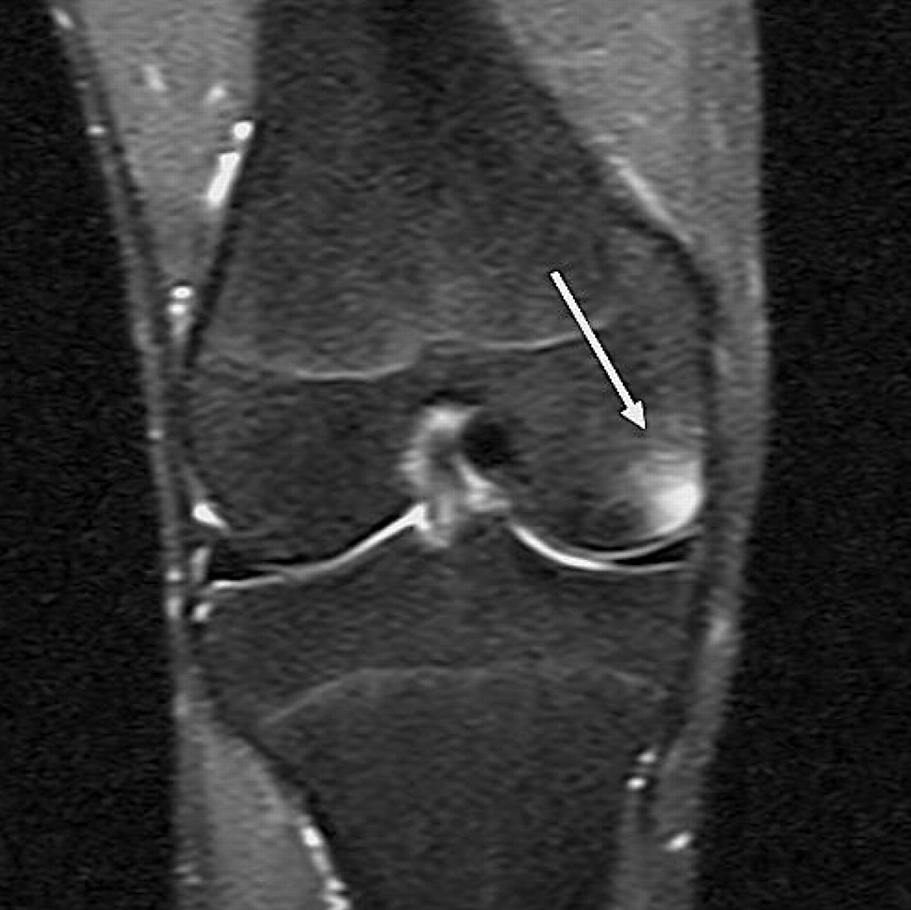
A teenage asymptomatic male swimmer. Bone contusion in the medial femoral condyle (arrow). Coronal STIR MR image (TR/TE, 4750/19).

A teenage asymptomatic male swimmer. Prefemoral fat pad edema and joint effusion (arrows). Sagittal STIR MR image (TR/TE, 4750/19).
In the control group, which comprised 14 asymptomatic individuals, 28 knees were examined. MRI revealed one or more abnormalities in only 9 (32.1%) of these 28 knees. The most frequent abnormality was infrapatellar fat pad edema, which was seen in 7 of the 28 knees (25%). The edema ran along the infrapatellar plica in 4 knees (14.2%) and was found in the superolateral portion of the infrapatellar fat pad in 4 knees (14.2%). One of the knees had edema in the two portions of the infrapatellar fat pad. Small popliteal cysts were found in two knees (7.1%). Differently from the study group, only one knee in the control group (3.5%) had bone marrow edema, found in the medial femoral condyle. No meniscus, ligament or cartilage abnormalities, and no joint effusions were found in the evaluation of the control group MRI exams.
The Fisher's exact test revealed a statistically significant difference in the overall frequency of abnormalities in the two groups (p=0.013) with a greater prevalence of abnormalities in the group of adolescent elite swimmers (table 1). The difference in frequency of infrapatellar fat pad edema, bone bruises, prefemoral fat pad edema and joint effusion between groups was statistically significant (p<0.05), and was much greater in the group of elite swimmers. The differences in other knee abnormalities were not statistically significant between the two groups of adolescent boys. Interobserver agreement was 0.82 (κ) for all evaluations.
Knee abnormalities – MRI findings
Discussion
The most important finding of the present study was that MRI of the knee joint revealed more imaging abnormalities in the asymptomatic elite swimmers than in the control group. Although swimming does not cause direct impact on the bone and ligament structures of the knee joint, it produces a chronically repetitive leg movement that may be associated with the imaging changes found in this study.12
Infrapatellar fat pad edema, found in more than half of the athletes (53.8%), may be related to the high rate of repeated knee joint extension movements during workout series, which can result in fat entrapment in the anterior femorotibial joint space. Similar findings have been shown in athletes who practice high-impact sports8 and in some prospective reports evaluating non-athletic individuals. These studies have revealed that positive MRI findings are not always associated with symptoms,25 although anterior knee pain may be related to MRI inflammatory signal changes along the infrapatellar plica26 or suprapatellar fat pad.27 28
Similarly, prefemoral signal changes, found in 19% of the swimmers, have already been described in previous reports focusing on athlete and non-athlete individuals.27 29 In these studies, MRI signal abnormalities were associated with fat signal, intermediate signal (similar intensity of muscle or cartilage) and fluid signal. They have suggested that the prefemoral fat pad may also be entrapped during knee extension in athletes along with the infrapatellar fat pad. Furthermore, when the signal corresponded to fat in this topography, there was no significant association with mass effect or knee pain, and this may be an explanation for this finding in asymptomatic patients in the present study.
Another frequent knee abnormality revealed by MRI was bone contusion, found in 26.9% of swimmers. This imaging finding has been described in athletes of several different sports11 23 30 and has not been fully associated with symptoms, not even in elite athletes. Major and Helms7 have found knee BMO in asymptomatic basketball players and have suggested that this abnormality may be assigned to the direct transmission of repetitive impact through articular cartilage to the underlying bone, which would cause this characteristic bone signal change. Another possible explanation for BMO in athletes has been reported by Vanhoenacker and Snoeckx30 who suggested that as a physiological response to repeated stress, a biomechanical change occurs as a result of training which leads to the development of edema in certain knee compartments. Regarding elite swimmers, the source of joint stress may be related to repetitive knee flexion and extension during leg movements in freestyle, butterfly and backstroke, and also associated with medial impaction during the whip kick movement in the breaststroke. The clinical meaning of BMO has been a focus of discussion since it was first described by MRI many years ago.31 Currently, there are still questions about the association between bone edema and knee pain.32 Major and Helms7 have raised a hypothesis that bone contusion in asymptomatic basketball players may be related to initial stress lesions at very early stages. Similarly, Lazzarini et al33 have used MRI in order to determine whether running can cause BMO and have suggested it may be a result of the sports activity itself. Kornaat et al11 also suggested BMO as a continuum injury that starts from physiologic response to biomechanical load and ends in stress fracture.
Joint effusions were revealed in 15.3% of swimmers. Previous reports have described minor joint effusions in asymptomatic subjects, who were not associated with sports practice or knee lesions.34 In athletes, it has been described as an MRI finding in asymptomatic individuals.23 24 Boks et al24 have described difficulty to determine which volume of joint effusion would be physiological or pathological. On the other hand, larger cut-off points have been correlated with knee joint lesions. Although joint effusion can be frequently related to an underlying lesion (eg, cartilage lesion, meniscal tear or ligament injury), no such abnormalities were detected in the performed MRIs.
In this study, the athletes had practiced all swimming styles in a similar frequency, duration and intensity of workouts, because they had not yet specialised in one style. This may be one of the limitations of this study, because it was not possible to correlate an association between swimming style and MRI abnormalities. The cross-sectional design and the relatively small number of evaluated athletes were other possible study limitations. Certainly, a longitudinal study with a larger number of athletes would allow further inferences. Another limitation was the low-field magnet used in the present study, which has slightly less sensitivity for detection of cartilage lesions compared to high-field units.20 Despite this, no cartilage lesion was found in any of the 56 scanned knees. To confirm these findings, a high-field magnet should be used in a subsequent study.
All evaluated athletes had regular check-ups by a physician in their swimming association. The observed imaging findings were described and reported to this physician in order to initiate a possible intervention.
In conclusion, the most prevalent abnormalities found in this study were infra- and suprapatellar fat pad edema, BMO and joint effusion. These results are consistent with previous studies in asymptomatic athletes of other sports.7,11,13,20,21,35,36 The high prevalence of positive imaging findings detected in the group of asymptomatic swimmers may correspond to possible benign changes or preclinical lesions, potentially deleterious in the future. To address these specific questions, longitudinal cohort studies should be conducted to better understand the significance of these early imaging abnormalities revealed by MRI.
What is already known on this topic
Most of the previous reports have used MRI to evaluate knee joint abnormalities of asymptomatic athletes in different sport modalities, such as soccer, basketball and gymnastic. To the best of our knowledge, no study has evaluated MRI abnormalities of knee joints in asymptomatic young elite swimmers.
What this study adds
MRI can reveal preclinical imaging abnormalities in the knee joints of young elite swimmers. This advanced imaging technique can predict and prevent possible deleterious injuries. However, a follow-up longitudinal study is necessary to better understand the significance of these early MRI abnormalities detected in this group of elite swimmers.
References
Footnotes
-
Competing interest None.
-
Patient consent Obtained.
-
Ethics approval This study was approved by the Ethics in Research Committee of the Hospital São Lucas of Pontifícia Universidade Católica do Rio Grande do Sul, Porto Alegre, Brazil.
-
Provenance and peer review Not commissioned; externally peer reviewed.