Article Text

Abstract
Background: The combined positioning of the trunk and knee in the coronal and sagittal planes during non-contact anterior cruciate ligament (ACL) injury has not been previously reported.
Hypothesis: During ACL injury female athletes demonstrate greater lateral trunk and knee abduction angles than ACL-injured male athletes and uninjured female athletes.
Design: Cross-section control-cohort design.
Methods: Analyses of still captures from 23 coronal (10 female and 7 male ACL-injured players and 6 female controls) or 28 sagittal plane videos performing similar landing and cutting tasks. Significance was set at p⩽0.05.
Results: Lateral trunk and knee abduction angles were higher in female compared to male athletes during ACL injury (p⩽0.05) and trended toward being greater than female controls (p = 0.16, 0.13, respectively). Female ACL-injured athletes showed less forward trunk lean than female controls (mean (SD) initial contact (IC): 1.6 (9.3)° vs 14.0 (7.3)°, p⩽0.01).
Conclusion: Female athletes landed with greater lateral trunk motion and knee abduction during ACL injury than did male athletes or control females during similar landing and cutting tasks.
Clinical relevance: Lateral trunk and knee abduction motion are important components of the ACL injury mechanism in female athletes as observed from video evidence of ACL injury.
Statistics from Altmetric.com
Adolescent and mature females who participate in pivoting and jumping sports suffer anterior cruciate ligament (ACL) injuries at a 2–10-fold greater rate than male athletes participating in the same high-risk cutting and landing sports.1–6 The combination of this greater susceptibility and a 10-fold increase in the female sports population since the inception of Title IX of the US Education Amendments act of 1972 (now known as the Patsy T Mink Equal Opportunity in Education Act) has resulted in a dramatic increase in the number of ACL injuries in females.5 In the USA, 100 000 to 250 000 ACL injuries occur each year.7 8 The costs exceed US $650 million annually in solely female high school and collegiate varsity athletics.9 Neuromuscular control deficits at the hip and trunk may contribute to decreased active neuromuscular control of the lower extremity (LE) that may lead to increased knee abduction loads and strain on the knee ligaments.10–13 In addition to the costs associated with ACL reconstruction and rehabilitation, there is a strong association between ACL injury and development of posttraumatic knee osteoarthritis, which also occurs with much greater incidence in females than males.14 15 It is estimated that between 50% and 100% of women with an ACL injury will show significant pain, functional limitations and radiographic signs of knee osteoarthritis within 12 to 20 years of the index injury.14 16
Knee abduction loads and neuromuscular control of the trunk both predict ACL injury risk with high sensitivity and specificity in female athletes. Knee abduction load predicted ACL injury risk with 78% sensitivity and 73% specificity.12 Trunk displacement, and specifically lateral trunk displacement, predicted risk of knee, knee ligament and ACL injuries with high sensitivity and specificity in female, but not male, athletes.17 A logistic regression model that incorporated lateral trunk motion predicted ACL injury risk in females with 83% sensitivity and 76% specificity, but did not predict knee or ACL injury risk in males. The mechanism of non-contact ACL injury may differ in females and males, especially with respect to the dynamic positioning of the knee, as females demonstrate greater valgus collapse of the lower extremity primarily in the coronal plane.18 Most ACL injuries in females occur by non-contact mechanisms during landing, deceleration and lateral pivoting.19 The mechanism of non-contact ACL injuries as observed on video has several common components in female athletes: high knee abduction, lateral trunk motion with the body shifted over the injured leg and the plantar surface of the foot fixed flat on the playing surface, displaced away from the centre of mass of the body and low knee flexion.18–21 Perturbation of the trunk, game or competitive situation and another player within close proximity are other common components of the mechanism.18 22
Trunk motion can influence knee abduction load through mechanical and neuromuscular mechanisms. If the trunk moves laterally, the ground reaction force (GRF) vector may move laterally and have a greater lever arm relative to the knee joint centre. This will directly increase the potential for knee abduction loading, especially if at the same time the magnitude of GRF increases due to unicompartmental (ie, lateral compartment) knee joint loading and/or increased inertial acceleration of the trunk or thigh segments during dynamic movement. Knee abduction torque places knee ligaments in the high slope (load) segment of their force-length curve and elicits knee pain in female athletes.23 24
The purposes of the present study were to determine the trunk and knee position of female and male athletes at the time of ACL injury, and to compare these results with those of uninjured control athletes. The tested hypothesis was that compared with female control athletes and male subjects, injured female subjects would show greater lateral trunk angles and greater knee abduction angles at landing after a jump, pivoting or after a sharp deceleration manoeuvre.
MATERIALS AND METHODS
Data collection
During a 12-year period (1995 to 2007), we requested from physicians, athletic trainers, patients and the National Basketball Association (NBA Entertainment (NBA and Womens NBA (WNBA) games)) videotapes of athletes captured during an ACL injury requiring reconstruction. A total of 70 such videotapes were collected. Our study was exempt from institutional review board approval. Criteria for inclusion of a video in our study were: (1) good quality, with the camera angle approximating a sagittal (lateral) or coronal (anterior or posterior) view of the athlete; (2) visualisation of the foot contacting the ground; (3) unobscured view of the athlete; and (4) no or minimal contact during the athletic manoeuvre. Minimal contact included being touched by an opponent, such as shoulder to shoulder contact during a rebound. Videotapes were excluded if the athlete was being tackled or pushed by an opponent or if there was any direct contact to the knee. For a more detailed account of these methods see Boden et al.22
In all, 23 injury videos met the criteria for this study: 10 female and 7 male ACL-injured players and 6 female controls performing similar landing and cutting tasks. We tabulated injury conditions such as type of sport, level of play, game or high-intensity practice situation, level of contact (none or minimal), activity being performed (vertical jump, broad jump, or deceleration), whether the player was on offence or defence, whether the subject was holding a ball and whether another player was in close proximity (being guarded or guarding another athlete).22
The same author selected and assessed videotapes of professional and collegiate basketball players (controls) performing similar decelerating or landing manoeuvres during game situations.22 Basketball was the sport of choice for the controls because of the availability of high-quality videos from professional and collegiate matches and because of the close proximity of the camera to the athletes.22
Video editing and analysis
The video recordings were edited using Adobe Premiere Pro (V 2.0, Adobe Systems, San Jose, California, USA) and deinterlaced to achieve a 30-Hz (frames/s) effective frame rate via Abode Photoshop (V CS2, Adobe Systems).22 Initial contact (IC) with ground was analysed on each video. The trunk and joint kinematic measures were performed from the video sequences using ImageJ software (http://rsb.info.nih.gov/ij/). Joint angles were analysed in five sequential frames (stored as TIFF files) in sagittal or coronal planes, starting with initial ground-foot contact (time 0). Therefore, the time sequences observed were at approximately, initial contact of the foot with the floor and 50 ms, 100 ms, 150 ms and 200 ms post contact. ImageJ was used to measure joint angles after drawing lines based on the landmarks described below.25 26 All measurements were performed by the same author for consistency.
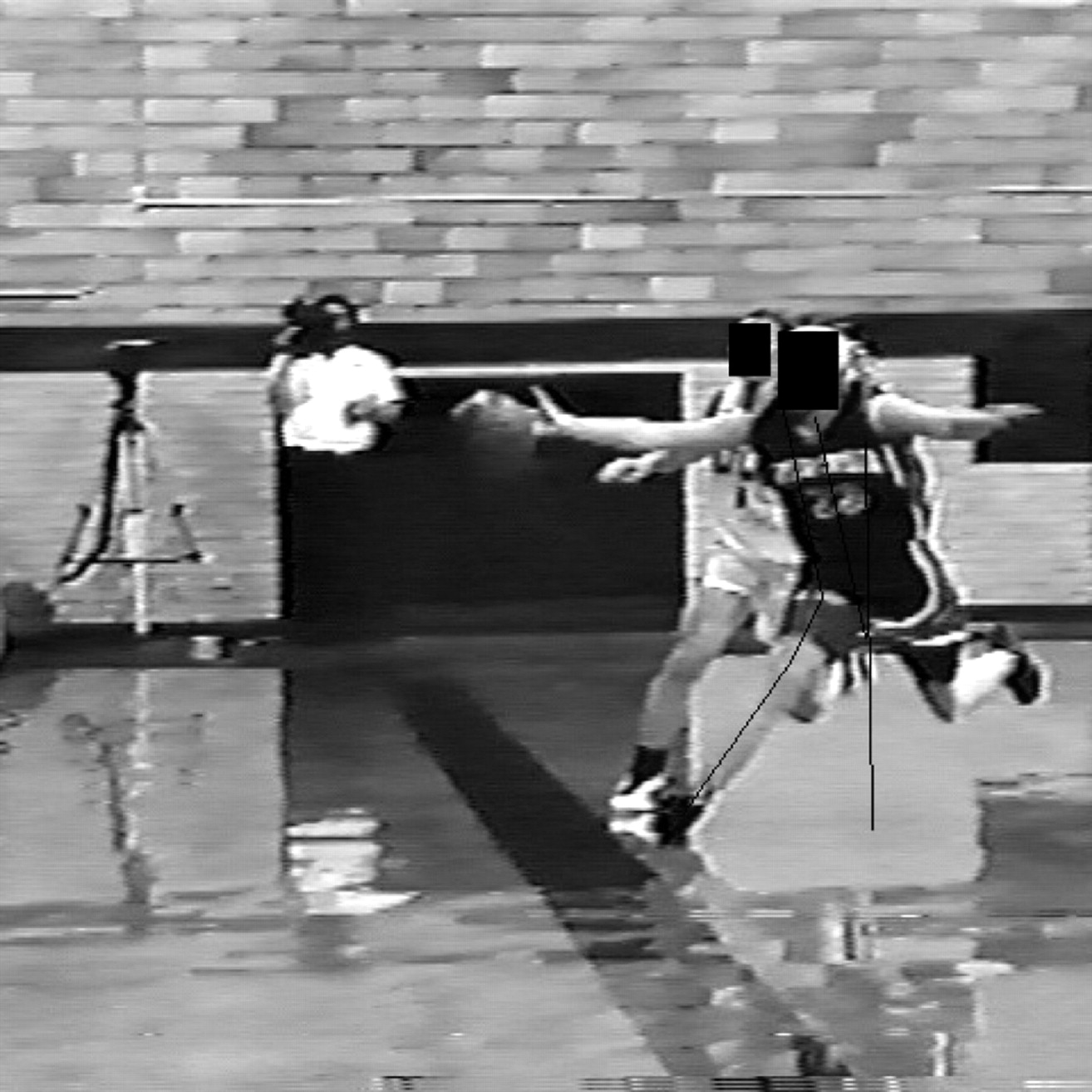
The trunk position was measured by connecting a line perpendicular to the floor and through the superior tip of the greater trochanter and a line drawn from the superior tip of the greater trochanter to the superior tip of the acromioclavicular joint (fig 1). For the anterior views the trunk lean angle was measured by connecting a line from the centre of the neck at the C7 level to the centre of the symphysis pubis and a line from the centre of the symphysis pubis perpendicular to the ground. For the posterior frames the trunk bend was measured by connecting a line from the centre of C7 to the centre of L4 and a line perpendicular to the ground and intersecting the centre of L4. The anterior and posterior views were analysed together to assess the coronal position. For a more detailed account of these methods see Boden et al.22

Still image of a female anterior cruciate ligament (ACL)-injured subject during injury (front player, no. 22) relative to a control player (behind her), demonstrating the association between lateral trunk motion and medial knee collapse in the injured subject, but not the control player (obscured view, not analysed). This is frame 1 (initial foot contact with ground) of the subject (no. 22) landing and shows the calculated angles and the combined lateral trunk motion and knee abduction.
Statistical analysis
All angle measurements were imported into Statview V 5.01 (SAS Institute, Cary, North Carolina, USA) for statistical analysis. Repeated measures analysis of variance (ANOVA) with Fisher protected least significant difference (PLSD) post hoc tests were performed to assess if there were statistically significant differences between female subjects and controls, and male and female ACL-injured subjects. Significance was set at p⩽0.05. Intraclass coefficients (ICCs) were calculated to assess the reproducibility of the angle measurements at each video frame sequence at three different times. The single rater repeated the measured videotape frames of 4 angles in a total of 10 subjects (2 angles/6 subjects; 2 angles/4 subjects). The estimated ICCs ranged from 0.32–0.99, with 18 of the 20 coefficients greater than 0.95.22
RESULTS
Lateral trunk angle
Figure 1 shows an ACL-injured subject demonstrating combined lateral trunk motion and knee abduction during ACL injury. The mean lateral trunk angle relative to the vertical was higher in female athletes during ACL injury than in male players during ACL injury (p = 0.02; fig 2) and trended toward being greater than female controls (p = 0.16) performing similar landing and cutting tasks across the five repeated measures frames assessed, or approximately 200 ms of landing (fig 3). These differences were significant between female and male ACL-injured subjects at IC (mean (SD) 11.1 (2)° vs −5.5 (9.5)°, p = 0.04) and trended toward being greater than female controls performing similar landing and cutting tasks (IC: 11.1 (1.2)° vs 4.2 (9.6)°, p = 0.29). The mean anterior–posterior trunk angle relative to vertical was not different in female athletes during ACL injury than in males (IC: 1.6 (9.3)° vs −6.7 (8.6)°, p = 0.20). In addition, female ACL-injured athletes demonstrated less forward trunk lean than female controls (IC: 1.6 (9.3)° vs 14.0 (7.3)°, p = 0.005).

Still image with coronal angles a male subject demonstrating the absence of an association between lateral trunk motion and medial knee collapse during anterior cruciate ligament (ACL) injury.

Coronal plane trunk angles (mean (standard error of the mean (SEM))) in female and male anterior cruciate ligament (ACL)-injured subjects and female controls.
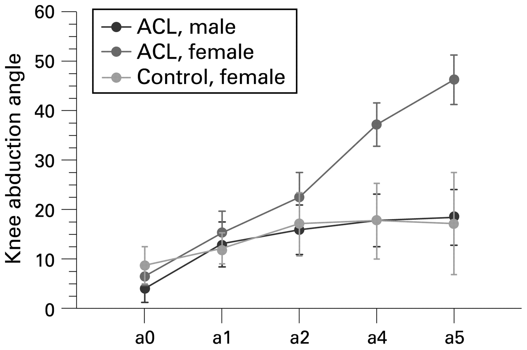
Knee abduction angle
Female ACL-injured subjects demonstrated significantly increased knee abduction during landing compared to male ACL-injured subjects and showed a trend toward more knee abduction than female controls (p⩽0.05; fig 4). After initial contact, the knee abduction moment remained relatively unchanged in the controls, but the female ACL-injured subjects showed progressively more knee abduction with each consecutive sequence (fig 4). The mean differences between subjects and controls for the third through fifth frames were significant (p⩽0.05). No significant differences were observed in knee flexion angle between subjects and controls across the five repeated measure frames. There were no significant differences observed in knee flexion between either female and male ACL-injured subjects or between female ACL-injured subjects and female controls.22

Coronal plane knee abduction angles (mean (standard error of the mean (SEM))) in female and male anterior cruciate ligament (ACL)-injured subjects and female controls.
DISCUSSION
We accepted the alternative hypothesis for the present study, namely that injured female subjects would show greater lateral trunk and greater knee abduction motion at landing compared to male injured subjects and uninjured female control athletes, since injured female subjects were observed to demonstrate greater lateral trunk and greater knee abduction angles than male subjects and female controls during landing injury (see figs 3 and 4 and Supplementary material). To our knowledge, there is no similar published data on trunk position in either descriptive or analytic studies of ACL injury. Trunk stability is related to the ability of the hip to control the trunk in response to forces generated from distal body segments as well as from unexpected perturbations.17 27 Deficits in neuromuscular control of the trunk during cutting and landing may lead to uncontrolled lateral trunk motion that may increase knee abduction motion and torque through mechanical (lateral GRF motion) and neuromuscular (increased hip adductor torque) mechanisms.11 12 Insufficient neuromuscular control of the trunk may increase strain on the ACL and lead to injury via either one or both of these mechanisms.10–13
Neuromuscular control of the hip is required to stabilise the trunk and pelvis. An external hip abduction moment created by the GRF moving lateral to the centre of the femoral head is counterbalanced internally by hip adductor torque. For example, during normal gait, with the lateral trunk motion that occurs in early stance, the hip adductors are activated in order to adduct the pelvis and move the trunk toward the midline.28 Females activate the hip musculature differently than males in response to sudden loading.29 Women adduct the hip more than men during low and high intensity activities. They begin descent in a more abducted knee position and remain in a more abducted alignment relative to men throughout a squat motion or during landing.30 31 Females also demonstrate more hip adduction than males during cutting.32 33 Increased hip adduction during dynamic motion and decreased hip muscle abductor strength and recruitment can increase knee load and injury risk. Ipsilateral trunk lean may be a sign of weak hip abductors as it moves the centre of mass closer to the stance limb to reduce demand on the weak abductors.27 34 During single leg landing and cutting, the entire body mass must be balanced over one lower extremity. Because the trunk comprises greater than half of the body’s mass, lateral trunk motion increases GRF and knee abduction load.34
Deficits in neuromuscular control of the trunk may contribute to lower extremity joint instability and injury.35 This may be due to a resulting decrease in active joint stiffness in women than men.36–38 Landing and cutting require high levels of neuromuscular control to maintain stability and performance.39 40 Dynamic stability of the knee is dependent upon accurate sensory input and appropriate motor responses to rapid changes in body position during cutting and landing.12 35 Neuromuscular control of the hip, trunk and knee is based on feedback control. The position and load of each segment is used to modify the descending movement commands.41 Impaired control of the hip and trunk can increase lower extremity injury. For example, abdominal muscle fatigue contributes to hamstring injuries.42 Subjects with ankle sprains had a delay in onset of gluteus maximus and medius activation.43 Female, but not male, athletes who suffered ankle injury had greater body sway prior to injury than uninjured controls.44
Movement biomechanics and lower extremity strength can be altered in females with neuromuscular training.45 Neuromuscular power can increase within 6 weeks of training and may result in decreases in peak impact forces and knee abduction moments.45–48 Observed changes in females may be greater than in males as their baseline neuromuscular performance levels are lower.49 If neuromuscular training can decrease ACL injury risk, it is likely that the mechanisms underlying increased risk are neuromuscular in nature.50 ACL injury risk may be reduced in trained females during landing and cutting.51 Elite female athletes show reductions in ACL injuries with neuromuscular training.52 These prospective studies indicate that neuromuscular training has the potential to decrease ACL injury rates in females.
Neuromuscular control of the trunk and lower extremity can be improved with neuromuscular training,12 45 47 53 54 which may increase coronal plane trunk and hip control in females.45 49 55 For example, during a drop-jump, a two-footed plyometric activity, post training results showed that lower extremity valgus was reduced at the hip. Conversely, during a single-leg landing task, the most significant modifications may occur at the knee.47 Therefore, the effects of training in the coronal plane are likely to be movement task specific.47 Increased coronal plane control at the hip and trunk may be necessary to reduce ACL injury risk.56–58 lower extremity coronal joint motions and torques linked to increased ACL injury risk are often correlated, indicating that control of knee load may require synergistic and antagonistic contribution from the trunk, hip and knee.12 Perturbation-enhanced training may increase trunk control and decrease knee abduction load in females.
The current video evidence of ACL injuries shows that the female trunk usually moves lateral to the ACL-injured limb as the knee abducts (fig 1 and Supplementary material), while this is not a common observation in males (fig 2). Trunk position and knee load may be mechanically linked, as lateral positioning of the trunk can create abduction loads at the knee.28 In the coronal plane, applying static equilibrium mechanics and neglecting the inertia of the body segments between hip and ground, if the GRF passes lateral to the centre of the head of the femur, an external hip abduction torque results.28 60 Interestingly, even alteration of arm position relative to the centreline of the body can increase the external knee abduction load by 29% to 60%.56 At the low knee flexion angles that are present during ACL injuries, the ACL, rather than the MCL, can be the primary restraint to knee abduction loads.56 61 62 Knee abduction load and ACL injury may be outcomes that result from an unstable, collapsing lower extremity column under axial load, caused by the GRF passing through the lateral knee compartment.
Females tend to utilise greater coronal plane control rather than a sagittal plane control strategy for the lower extremity.63–66 They tend to utilise a “hip strategy” for single leg control and balance during landing and cutting.30 67 For example, coronal plane excursions are greater and more rapid at the hip and knee during walking in females.59 The knee functions optimally as a sagittal plane hinge, not a coronal plane hinge or ball and socket joint, as the large muscles of the lower extremity that limit coronal plane trunk, hip and knee motion or torque absorb and dissipate force most effectively and efficiently in the sagittal, rather than coronal plane.68
A coronal plane, quadriceps dominant (ie, flat footed and knee abducted) landing strategy likely leads to higher ground reaction and ACL injury risk in female athletes. Females that land with knee abduction are at increased risk for ACL injury.7 12 A combined flat foot and abducted knee position likely result in high axial ground reaction forces in the lateral compartment of the knee joint. With combined knee abduction and flat foot, the ground reaction force cannot be effectively absorbed by the prime movers of the lower extremity.69 If there is an axial ground reaction force on the lateral joint, this may lead to internal rotation of the tibia on the femur, which increases strain on the ACL.13
Previous studies of ACL injuries based on videotape analysis have relied on visual inspection to determine joint positions.19 70 71 Visual inspection has poor accuracy and precision for joint angle measurement.72 In a study assessing the accuracy of the visual inspection technique, the mean error for knee flexion was 19 degrees, while the standard deviation between the observers for hip flexion was 18 degrees on average.72 We used a digital measuring tool to define joint position more accurately and more quantitative.
The present observations show that lateral trunk rotation to the ipsilateral side likely increases valgus, axial and/or compressive forces on the lateral side of the knee joint, lowering the threshold for ACL injury. These findings agree with those of Krosshaug et al that female athletes showed greater knee abduction position (“valgus collapse”) after and possibly during ACL injury than do their male counterparts. There was also a trend in our female subjects toward more knee abduction after landing than was shown by male subjects; this finding may indicate that there is more inherent knee abduction in females during landing than in males, which concurs with the data of Hewett et al.12 Hewett et al7 12 showed that a landing pattern with knee abduction is a risk for ACL injury, which may explain the higher incidence of non-contact ACL injury in women than in men. It is possible that with knee abduction, the axial forces are greater on the lateral side of the knee than on the medial side, further enhancing the lateral compressive forces and allowing for a greater internal rotation component to the injury. In addition, with knee abduction, the ligaments on the lateral side of the knee are relaxed while the medial side tightens, allowing the lateral side to shift anteriorly and rotate.69 Matsumoto showed that, with a valgus torque, the axis of the pivot shift is located at the medial collateral ligament.69 If the medial collateral ligament is taut, the movement of the medial side of the tibia is limited.69 In contrast, there is an axial or compressive force on the lateral joint. The combination of medial and lateral compartment forces may lead to internal rotation of the tibia on the femur, which can dramatically increase the strain on the ACL.13
Increased hip abductor muscle recruitment and strength likely has a direct effect on the knee abduction loading of the ACL during cutting and landing. Though ACL injuries likely occur too quickly (likely under 100 ms) for reflexive muscular activation (greater than 100 ms), athletes can adopt preparatory muscle recruitment and movement patterns that reduce the probability of injuries caused by unexpected perturbations.24 73 74 We hypothesise that decreased neuromuscular control of the trunk leads to increased joint load (knee abduction moment) via lateral motion of the GRF and results in increased ACL injury risk in female athletes (figs 1 and 3). In addition, the sagittal trunk observations were that the injured female subjects had less trunk flexion than female controls. This may place their trunk in line with the leg to increase valgus and/or the axial forces. However, decreased flexion at trunk did not appear to alter flexion at other lower extremity joints.
Potential limitations
There were several limitations to our study. We had a relatively small sample size of videotapes, which were collected as a convenience sample, for each camera angle. These videotapes may not be representative of all non-contact ACL injury mechanisms, but the observed motions likely represent some of the most common non-contact or minimal contact mechanisms of ACL injury. We were unable to determine the exact moment at which the ACL injury occurred. However, by measuring several consecutive frames in which the knee was deforming abnormally (compared to the knee of a control) and the athlete fell to the ground and grabbed the knee, it is likely that the injury occurred within the five measured frames. In addition, although such conditions cannot be matched perfectly in different groups, even in a laboratory setting, the descriptive findings for our subjects and controls were fairly evenly matched. Because measuring knee abduction in the coronal plane does not account for rotation of the leg (internal rotation of the femur and external rotation of the tibia), our coronal knee abduction angles may not be pure knee abduction but, rather, a combination of knee abduction, internal rotation of the femur and external rotation of the tibia. This problem may be compounded by the fact that the body may be rotating but the camera is being held still. Unfortunately, with a two-dimensional analysis, we were unable to separate these components of motion. Future studies analysing ACL injury videotapes where the injury was captured from more than one camera angle may be able to provide a more accurate three-dimensional assessment of the various components.75
There were also several potential limitations in our technical analysis: possible difficulties with identifying anatomic landmarks in clothed individuals with no markers, camera angle variability that may not have captured all individuals in a perfect sagittal or coronal plane, possible microsecond differences in the timing of the first sequence picked as the foot touched the ground to the point of injury and the limitations of two-dimensional analysis. However, this computerised technique of angle measurements is a considerable improvement over previous descriptive studies based purely on visual estimates of joint position. Despite these limitations, to our knowledge ours is the first study to analyse trunk position in videotaped ACL injuries with a group of controls for comparison. Kinematic analysis with two or more synchronised camera views would provide more accurate data and data not yet recorded in the literature.75
Summary and conclusions
This study may advance the understanding of the mechanisms and prevention of ACL injuries in female athletes, who are at a 2–10-fold increased risk of ACL injury than males. Our objectives are to determine the mechanisms by which female athletes become more susceptible to ACL injury and to optimise the effectiveness of interventions designed to prevent ACL injuries. Specifically, this study was directed towards understanding and answering the question of whether increased lateral sway of the trunk underlies increased abduction loading of the knee joint during ACL injury in female athletes. The present evidence supports this theory (see red arrows in Supplementary material). This information may enhance the efficacy of ACL prevention programs. Prophylactic intervention for ACL injury could prevent a significant percentage of the 100 000 to 250 000 injuries that occur each year in the USA.7 8 Reduction of female injury rates from fivefold greater to equal male injury rates would potentially allow females annually to continue the health benefits of sports participation and avoid the long-term complications of osteoarthritis, which occurs with a 10-fold to 100-fold greater incidence in ACL-injured than in uninjured athletes.14 16
REFERENCES
Footnotes
Competing interests: None.
Funding: This work was supported in part by NIH/NIAMS grants R01 AR049735, R01 AR05563 and R01 AR056259 (TEH).
Ethics approval: Ethics approval was obtained.
▸ Additional data (Supplementary figure 1) are published online only at http://bjsm.bmj.com/content/vol43/issue6