Article Text

Abstract
Background Insertional Achilles tendinopathy is well known to be difficult to treat, especially when there is intra-tendinous bone pathology. This study is a case series on patients with chronic insertional Achilles tendon pain and major intra-tendinous bony pathology together with bursa and tendon pathology, treated with excision of the subcutaneous bursa alone.
Methods Eleven patients (eight men and three women) with a mean age of 44 years (range 24–62) and a chronic (>6 months) painful condition from altogether 15 Achilles tendon insertions were included. In all patients, ultrasound examination showed intra-tendinous bone pathology together with pathology in the tendon and subcutaneous bursa, and all were surgically treated with an open excision of the whole subcutaneous bursa alone. This was followed by full weight-bearing walking in a shoe with open heel for 6 weeks.
Results At follow-up 21 (median, range 12–108) months after surgery, 9/11 patients (12/15 tendons) were satisfied with the result of the operation and 10/11 (13/15 tendons) were back in their previous sport and recreational activities. The median VISA-A score had improved from 41 (range 0–52) to 91 (range 33–100) (p<0.01).
Conclusion In patients with chronic painful insertional Achilles tendinopathy with intra-tendinous bone pathology, tendon and bursa pathology, open removal of the subcutaneous bursa alone can relieve the pain and allow for Achilles tendon loading activities. The results in this case series highlight the need for more studies on the pain mechanisms in insertional Achilles tendinopathy and the need for randomised studies to strengthen the conclusions.
Level of evidence IV Case series.
- Achilles
- tendon
- tendinopathy
- lower limb surgery
This is an open access article distributed in accordance with the Creative Commons Attribution Non Commercial (CC BY-NC 4.0) license, which permits others to distribute, remix, adapt, build upon this work non-commercially, and license their derivative works on different terms, provided the original work is properly cited, appropriate credit is given, any changes made indicated, and the use is non-commercial. See: http://creativecommons.org/licenses/by-nc/4.0/.
Statistics from Altmetric.com
What are the new findings?
Open removal of the subcutaneous bursa alone can relieve the pain in patients with chronic painful insertional Achilles tendinopathy with intra-tendinous bone pathology.
Despite the presence of severe bone pathology in the Achilles tendon insertion, pain may mainly originate from the subcutaneous bursa.
Introduction
Insertional Achilles tendinopathy is difficult to treat, and its pathogenesis has not been fully elucidated yet.1–3 It often includes pathology in multiple tissues, such as bursa, bone and tendon tissue, and the source of pain can be difficult to diagnose. Intra-tendinous bone formation is considered especially difficult to treat. Non-invasive conservative treatment is first line.2–4
However, when there is major intra-tendinous bone pathology involved, in clinical practice surgery is often warranted. Surgical techniques5–18 include procedures such as tendon detachment–bone removal–tendon re-attachment and calcaneo-osteotomy. Bone removal via longitudinal tenotomy is an alternative when there is minor bone pathology. Despite promising results in several studies,6 7 19 the rehabilitation period is long and includes an initial period with immobilisation.20
Traditionally, these surgical methods do not specifically focus on removal of the subcutaneous bursa. Recent research on innervation patterns has shown the subcutaneous bursa to be the most richly innervated tissue in patients with severe pathology in the Achilles tendon insertion.21 In this case series, we therefore evaluated patients with chronic painful insertional Achilles tendinopathy, having ultrasound-verified pathology in the subcutaneous bursa and tendon together with intra-tendinous bone pathology, where treatment consisted of excision of the subcutaneous bursa alone.
Materials and methods
Patients
Eleven consecutive patients (eight men, three women), mean age of 44 years (range 24–62), with chronic (>6 months) painful insertional Achilles tendinopathy in altogether 15 Achilles insertions, were included. All patients had tried conservative treatment; for example, rest, heel lift, concentric and eccentric loading regimens, non-steroidal anti-inflammatory drugs, injections (cortisone, Traumeel, platelet-rich plasma) and ultrasound (US)+Doppler-guided sclerosing polidocanol injections. None of the patients had inflammatory conditions known to involve tendons. All patients in the current study were active individuals, one triathlete, eight joggers and two walkers.
Clinical examination
There was a widened heel profile, including a thickened subcutaneous bursa with diffuse local tenderness. The distal Achilles tendon was thickened. Resting tonus and range of movement was normal. There were no clinical signs of a bony prominence in the region for upper calcaneus (Haglund deformity) and no obvious soft-tissue prominence in the region for retrocalcaneal bursa (retrocalcaneal bursa enlargement).
Dynamic US and Doppler examination showed a thick subcutaneous bursae with fluid-rich islands. Regions of high blood flow were seen in the bursal walls (figure 1). There was distal Achilles tendinopathy and intra-tendinous bone pathology (bone bridges, bone spurs, loose bone).

Longitudinal greyscale ultrasound+colour Doppler view: high blood flow in the subcutaneous bursa overlaying the bone formations in the distal tendinopathic Achilles.
For diagnostic evaluation, a US-guided injection of a local anaesthetic (2–4 mL of xylocaine+Epinephrine) into the subcutaneous bursa was used. If the patients were then pain-free during provocation with Achilles tendon loading activities, this indicated that subcutaneous bursa removal alone could be a sufficient treatment.
Surgical procedure
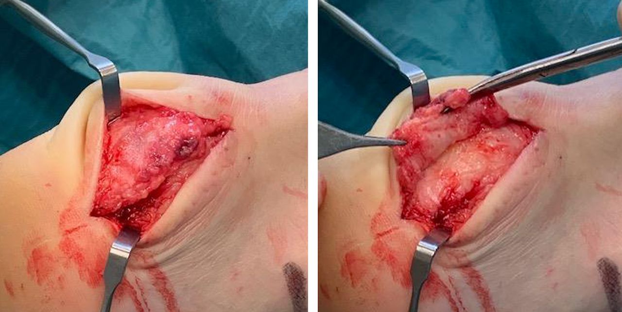
All patients were in local anaesthesia surgically treated with an open excision of the whole subcutaneous bursa alone. Via a longitudinal lateral incision in the Achilles tendon insertion, the subcutaneous bursa was identified and carefully released from the skin and tendon side, all the way over to the medial side, before being removed (figure 2). Often, the bursa was in regions coalesced with the tendon, including islands of fatty infiltration. Hemostasis was established and flush administered before closure. Bandage was from toes to below the knee, with local compression over the heel. For patients with bilateral condition, both sides were operated at the same time. Postoperative rehabilitation steps are described in table 1.
Overview of postoperative rehabilitation steps
Surgical removal of the subcutaneous bursa.
Follow-up
The patients were contacted via telephone or email. All patients answered a questionnaire including questions about “satisfied/not satisfied”, “pain/no pain”, “returned to previous activity/not returned to previous activity”, “skin problems (sensitive skin?)” and filled in VISA-A scores.
Ethical consideration
Studies on surgical treatment of Achilles tendinopathy were approved by the local ethics committee (Umeå University, Sweden). All patients signed an informed consent.
Patient and public involvement
Patients and public were not actively involved in this research study.
Statistics
SPSS was used to analyse the data (SPSS, Chicago, Illinois, USA). Normal distribution was tested using a Kolmogorov-Smirnov test. For the comparison of pre-VISA-A and post-VISA-A scores, a Wilcoxon test was used. Significance level was set to p value <0.05.
Results
All patients answered via a telephone call or mail contact. VISA-A scores were obtained from 9/11 patients (13/15 tendons). One patient had moved to another country, and one answered the telephone interview but did not send back the questionnaire and VISA-A scores.
At follow-up 21 months (median, range 12–108) after surgery, 9/11 patients (12/15 tendons) were satisfied with the results and 10/11 patients (13/15 tendons) were back in previous sport and recreational activities. The VISA-A score (median) had improved from 32 (range 0–52) to 91 (range 33–100) (p<0.01). Two patients were not satisfied. One patient operated on bilaterally had remaining pain at the back of both heels (VISA-A was 33 on the left side and 56 on the right side). The other patient was diagnosed with midportion Achilles tendinopathy, had minor pain (the VISA-A score was 80) and could participate in his previous recreational activities.
Complications: All patients reported varying degrees of a temporarily decreased touch sensibility in the skin at the back of the heel. The size of these regions varied. No patient reported this as a negative phenomenon, instead they were happy to not anymore suffer from intensive touch pain. In three patients, there were minor skin blisters during the first 6–10 weeks after surgery.
Discussion
In this case series on patients with chronic painful insertional Achilles tendinopathy, having ultrasound-verified pathology in bursa and tendon together with intra-tendinous bone pathology, excision of the subcutaneous bursa alone showed in almost all patients good clinical results at 21 months of follow-up.
Treatment of chronic painful insertional Achilles tendinopathy is challenging. When there is pathology in multiple tissues, it is difficult to identify the source for pain. For patients with intra-tendinous bone pathology and failed conservative treatments, major surgery is often instituted.19 Tendon detachment–bone removal–tendon re-attachment,2 3 or bone removal via longitudinal tenotomy,6 followed by initial immobilisation and long rehabilitation periods,20 is commonly used. The results vary, and the need for a long low-loading rehabilitation period is not optimal for any patient. Traditionally, the focus has been on removal of bone (spur, bone bridge, loose bone) and not the subcutaneous bursa. This methodology is likely based on the theory that the sometimes occurring major bone formations, or prominent loose bone, is the main source of pain.
Interestingly, in recent research on innervation patterns in patients with insertional Achilles tendinopathy, immunohistochemical analyses have shown that most nerves were found in the subcutaneous bursa.21 In that study, biopsies from the subcutaneous bursa, the retro-calcaneal bursa, the upper calcaneus and the ventral side of the distal Achilles were taken from patients having US-verified pathology in all these tissues.22 Thus, it is likely that the subcutaneous bursa is involved in the pain in this condition. Therefore, we routinely use diagnostic US-guided injections into the subcutaneous bursa alone, followed by provocative Achilles tendon loading activity. When patients are pain-free during provocation, it is likely that removal of the subcutaneous bursa alone (without addressing the neighbouring tissues) can give good treatment results. In a single case observation on an international high-level triathlete, there was a very good clinical result with only subcutaneous bursa removal despite major intra-tendinous bone pathology.23
To the best of our knowledge, there are no previous studies in chronic painful insertional Achilles tendinopathy that have focused only on removal of the subcutaneous bursa. Therefore, we used a preliminary study design to examine the effects of this specific treatment model.
There are advantages for the patients if removal of the subcutaneous bursa alone is enough to relieve the pain from this condition. The rehabilitation period is short and there is no need for total immobilisation. For athletes, it is often possible to stay in training, with some modifications. Furthermore, the operation is done in local anaesthesia, and possible risks with general, spinal and epidural anaesthesia can be avoided.
A possible disadvantage might be that in some patients, in the current material the skin quality posterior on the heel was not optimal (the skin sensibility was lowered and it was easier to get blisters) after surgery. This highlights the importance to protect the skin posterior on the heel the first 6 weeks after surgery, and for a few patients awareness seems to be needed also later on.
The patients in the current study were all active individuals performing endurance sport activities (triathlon, jogging) or at least regular longer distance walking. Ten patients were back in their Achilles tendon loading activities at desired level after surgery, indicating that the majority of pain they suffered was derived from the subcutaneous bursa.
A limitation is the relatively small patient material. Larger materials and randomised studies comparing bursectomy alone with procedures such as involving tendon detachment, bone removal and tendon re-attachment are needed.
For this condition that often involves pathology in multiple different tissues, it likely varies from what tissue the pain comes from. Given that the subcutaneous bursa has been shown to be the most richly innervated tissue in patients that have combined pathology in the bursae, tendon and bone, it seems logical to include diagnostic injection of local anaesthesia into the subcutaneous bursa as part of the preoperative evaluation. If the injection temporarily cures the pain, because of the major difference in the surgical treatment and postoperative rehabilitation, it can be recommended to first try the less invasive subcutaneous bursa removal alone procedure.
In conclusion, despite the presence of major bone pathology in the Achilles tendon insertion, pain may mainly originate in the subcutaneous bursa, a structure often ignored by traditional operations. Diagnostic injections can be used to verify pain from the subcutaneous bursa, and removal of the subcutaneous bursa alone is a treatment alternative. The results in this case series highlight the need for more studies on the pain mechanisms in insertional Achilles tendinopathy, and the need for randomised studies to strengthen the conclusions.
Acknowledgments
The authors would like to thank all the patients for their willingness to participate in this study.
References
Footnotes
Contributors HA performed all the surgeries and clinical examinations. CS performed the data analysis. Both authors were involved in the design of the study, the data collection and manuscript writing. The manuscript was finally approved by both authors.
Funding The authors have not declared a specific grant for this research from any funding agency in the public, commercial or not-for-profit sectors.
Competing interests None declared.
Patient consent for publication Not required.
Provenance and peer review Not commissioned; externally peer reviewed.
Data availability statement Data are available upon request.