Article Text

Abstract
Objective This study evaluates the intra-rater and inter-rater reliability of the MyotonPRO and its construct validity for the assessment of Achilles tendon stiffness.
Design Reliability and construct validity study.
Methods Forty healthy participants were assessed using the MyotonPRO by two raters on two different occasions. Tendon was evaluated in three different positions (relaxed, 0° plantarflexion and standing) and during different isometric contractions (range 0–3 kg). Reliability was calculated using intraclass correlation coefficient (ICC and 95% CI) standard error of measurement and minimal detectable change. Construct validity was evaluated between the different positions and the different contraction intensities using Friedman test.
Results Intra-rater reliability was very high ICC2,k 0.87–0.98. The reliability of the 0.5 kg contraction was moderate with an ICC2,k of 0.59. Inter-rater reliability ranged from high to very high with an ICC2,k of 0.76–0.86. The reliability of the 0.5 kg, 1 kg contraction and the standing position was moderate with an ICC2,k of 0.55, 0.54 and 0.56 respectively. Inter-session reliability ranged from high to very high with an ICC2,k of 0.70–0.89. The reliability of the 0.5 kg contraction was moderate with an ICC2,k of 0.54. Construct validity was demonstrated between different contraction levels and different positions.
Conclusion MyotonPRO is a reliable tool for the evaluation of Achilles tendon stiffness during different contraction levels and in different positions. Construct validity was supported by changes of tendon stiffness during the explored conditions. MyotonPRO can be implemented, as a ready to use device, in the evaluation of tendon tissue mechanical properties.
- ankle
- tendon
- achilles
This is an open access article distributed in accordance with the Creative Commons Attribution Non Commercial (CC BY-NC 4.0) license, which permits others to distribute, remix, adapt, build upon this work non-commercially, and license their derivative works on different terms, provided the original work is properly cited, appropriate credit is given, any changes made indicated, and the use is non-commercial. See: http://creativecommons.org/licenses/by-nc/4.0/.
Statistics from Altmetric.com
Summary box
Achilles tendon stiffness can be measured with MyotonPRO. High intra-rater, inter-rater and inter-session reliability has been found in different position and during isometric contractions.
The interest in measuring tendon stiffness is growing for its clinical and research relevance. A reliable and ready to use device has been presented in this study. MyotonPRO could be implemented in the evaluation of Achilles tendon mechanical properties.
Stiffness of healthy Achilles tendons has been assessed, reliability in patient with tendinopathies could be different.
Introduction
The Achilles tendon (AT) is one of the most commonly injured structures in the lower limb, both in athletes and the general population.1 2 Tendinopathy of AT can alter the mechanical, material and morphological properties of the tendon structure with a decrease of stiffness and young modulus and an increase of tendon cross-sectional area and diameter.3 4
Healthy tendon is highly responsiveness to mechanical loading.5 Adaptation of tendon has been shown following different loading protocols, and the intensity of the exercise (ie, high loading magnitude) and the intervention duration (>12 weeks) appear to contribute the most to changes in the mechanical properties of the tendon. The change in tendon stiffness appears to be largely due to adaptations in the material rather than morphological properties of the tendon.5 That is, early changes in young modulus is apparent following loading protocols whereas changes in tendon hypertrophy appear as longer-term adaptations to mechanical loading.6
Tendon responses to loading, unloading and ageing have been evaluated in a recent review revealing adaptability of the tendon tissue with an increase in stiffness when loaded and reduced stiffness when unloaded.3 Nevertheless, conflicting results has been reported when evaluating the in vivo effects of ageing on tendon stiffness, with some authors suggesting a decrease in modulus3 7–11 and others reporting unchanged tendon stiffness with ageing.12 13
Given the clinical and research interest in evaluate tendon stiffness as a parameter to describe tendon behaviour and tendon health, different measurement techniques have been developed. Once recent, non-invasive, ready to use technology is the MyotonPRO (Muomeetria, Tallinn, Estonia). This device is a handheld myotonometer producing a mechanical impulse to the skin overlying the target structure. The oscillation of the tissues underneath the probe permits calculation of the viscoelastic properties of the tissue.14–16 One parameter that can be extracted with this device is dynamic stiffness (N/m), which has been used in different studies to characterise the behaviour of different musculoskeletal tissues. This device has recently been used to evaluate tendon stiffness17–20 with a recent work showing decreased stiffness over the tendinopathic region of the tendon in people with Achilles tendinopathy and a significant difference in tendon stiffness between weight-bearing and non-weight-bearing positions.
The reliability of the MyotonPRO has been demonstrated for the evaluation of muscle stiffness,15 21–23 however, the reliability of MyotonPRO for measuring AT stiffness requires further investigation,17 18 24 25 especially regarding the capability of this device to measure the tendon in different conditions. In contrast to muscle, values of AT stiffness are extremely high and even higher when tested under load.19 Being able to quantify AT mechanical properties with a ready to use and affordable device which provides a reliable and valid evaluation of the tendon under different conditions, would provide new opportunities to characterise the mechanical behaviour of the AT, for example, in people with Achilles tendinopathy or following an intervention.
This study evaluates the intra-rater and inter-rater reliability of using the MyotonPRO to measure stiffness of asymptomatic AT in different position and during increasing load and to determine the construct validity of this device for measuring tendon stiffness.
Methods
The study was conducted between January and July 2019 at the University of Applied Sciences and Arts of Southern Switzerland. The study reporting adheres to the Guidelines for Reporting Reliability and Agreement Studies.26
A convenience sample of 40 healthy participants (18 women, 22 men, aged 33.8±14.2 years) were prospectively recruited trough public announcement at the University. Their mean (±SD) height and weight were 172.2 (±8.2) cm and 71.7 (±12) kg. Health-related physical activity was monitored using the International Physical Activity Questionnaires Short form (IPAQ)27; 23 participants were categorised as high, 15 as moderate and 2 as having low physical activity levels. Participants were excluded if they had a history of tendon or foot injury or surgery; connective tissue, metabolic or endocrine disease. Furthermore, participants with systemic inflammatory disorders, rheumatoid arthritis, spondyloartropathy or hypercholesterolaemia were excluded.
Sample size was estimated according to Walter et al28 using the following parameters: two replicates, p0=0.6, p1=0.80, α=0.05 and β=0.2. The optimal sample size was 39.1, and we rounded up to 40 participants.
Procedures
The AT mechanical properties were randomly analysed by two independent operators who were both physiotherapists with a minimum of 4 hours of training with the MyotonPRO prior to the study. Two measurement sessions were planned within 7 days. During the first session, the two operators consecutively performed the measures in order to evaluate the intra-rater and inter-rater reliability. The second session was used to evaluate inter-session reliability.
Testing location and room temperature (21°C–23°C) was kept constant across sessions. Participants were asked to maintain the same activity level between the two testing days.
The resistance of the soft tissues to the deformation induced by the MyotonPRO is calculated from an acceleration graph representing the damped natural oscillation of the tissues. Dynamic stiffness, calculated in N/m, is computed by multiplying the maximum acceleration of the oscillation graph to the mass of the probe divided by the maximum displacement of the tissue; higher values represent a stiffer tissue.
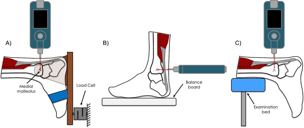
Both the left and right AT were evaluated at the most central point of the tendon following a trajectory from the most central component of the medial malleolus as this point has been suggested as the most prevalent region for tendinopathy.29 Furthermore, a previous study using MyotonPRO to evaluate stiffness of the AT in people with Achilles tendinopathy and asymptomatic controls showed that changes in mechanical parameters are more likely to be detected from 8 to 12 cm from the plantar aspect of the heel, corresponding to the free tendon part.19
MyotonPRO was setup with a multiscan sequence of 5 impulses (1 s apart), a short duration (15 ms) impulse involving minimal mechanical force with a precompression of 0.18 N and an impulse of 0.40 (total=0.58 n) was exerted on the tendon.
Measurements ware taken in three different positions; standing, a relaxed position and during isometric contractions of the plantar flexors of increasing intensities, in order to evaluate construct validity (figure 1). As the triceps surae contracts to increase plantarflexor force, the passive tension within the tendon increases as shown in previous studies.30 Since tendon stiffness and tendon displacement are directly related to plantarflexor strength31 and ankle angle,32 myonotometry values are expected to increase with increasing muscle contraction and with increased plantarflexion. To illustrate this construct, stiffness of the AT was compared across the five different contraction levels and between the three different positions. Testing in the three different positions as well as the sequence of the isometric contractions were randomised using a random number table.

Schematic of the experimental setup. (A) Isometric contractions: the foot was securely attached to a dynamometer with a wooden board at a fixed angle of 0° of dorsiflexion. The force measured by the load cell was proportional to the torque exerted at the ankle level. (B) Standing position: participants were asked to stand on a Wii balance board (Nintendo, Kyoto, Japan) with the centre of mass centred on the centre of board with the feet equally spaced. (C) Relaxed position: the feet were hanging freely over the edge of the examination bed. MyotonPRO was applied to the most central point of the tendon at the level of the medial malleolus.
Isometric contractions
The participants were positioned in prone with the foot secured within a dynamometer with the ankle in 0° of dorsiflexion/plantarflexion. Different intensities of isometric contractions of the plantar flexors were performed (0, 0.5, 1, 2 and 3 kg). The torque at the ankle joint was evaluated using a dynamometer connected to a force sensor operating in a 0–100 n range (CCT Transducers, Turin, Italy). The force sensor was attached to a wooden board connected to a hinge that enabled natural movement of the ankle joint in dorsiflexion and plantarflexion (figure 1A). The board was then fixed in order to permit isometric contractions only at a defined angle of 0° dorsiflexion. The force detected by the sensor was proportional to the torque exerted at the ankle level. Force signals were amplified using MISO-II (OT Bioelettronica, Turin, Italy) and visual feedback was provided to the participants.
A practice phase prior to the recording was conducted by asking the patient to familiarise with the visual feedback and perform contractions with the instruction to ‘push on the board as if you want to reach a tip-toe position’. During this practice phase, the lift of the heel was controlled. In order to prevent the detachment of the heel from the board, the foot and ankle were tightly secured in the dynamometer with two belts.
Standing
For the analysis in the standing position, participants were asked to stand on a Wii-Balance board (Nintendo, Kyoto, Japan) with the centre of mas on the middle of the board with the feet equally spaced (figure 1B). Visual feedback was provided by the BrainBLOX Software (University of Colorado, Boulder, Colorado, USA) to the participant in order to ensure that the centre of mass remained in the same position. Participants were asked to rest their hands on a fixed box which ensured the stability of the standing position and avoided oscillation of the centre of mass during the measurement that could affect the tension of the triceps surae muscle-tendon complex.
Relaxed position
The measurements in the relaxed position were performed with the participants in prone on an examination bed and the feet hanging freely over the edge (figure 1C). The participants were instructed not to move their feet during the measurement.
Statistical analysis
Statistical Package for the Social Sciences (SPSS V.24) was used to perform statistical analysis. Descriptive statistics (mean±SD) were used to describe the demographic characteristics of the sample. The distribution of the values was assessed using the Shapiro-Wilk test. Given the non-normal distribution of the data, non parametric, independent samples, Mann-Whitney test was used to assess whether there were significant differences in AT mechanical properties between men and women, between participants with different physical activity levels based on their IPAQ score.
Intra-rater and inter-rater reliability was assessed using intraclass correlation coefficient (ICC2,k) and the absolute reliability was calculated using standard error of measurement (SEM) and minimal detectable change (MDC).33
Furthermore, a cut-off point of ICC >0.6 was used to consider the clinical relevance of the measurements.34 35
Construct validity was tested analysing the correlations between the five different contraction levels and the three different positions using Friedman test with post hoc pairwise comparison Bonferroni correction was applied and statistical significance was set to α=0.05. Data are presented as median and IQR.
Patient and public involvement statement
This research was done without patient involvement. Patients were not invited to comment on the study design and were not consulted to develop patient relevant outcomes or interpret the results. Patients were not invited to contribute to the writing or editing of this document for readability or accuracy.
Results
No significant difference in AT stiffness was observed between men and women (p=0.6) or between participants with different physical activity levels based on their IPAQ score (p=0.9), therefore, the data were pooled and further analysed across the sample. Table 1 reports the descriptive statistics for the sample in the different conditions.
Descriptive statistics
A total of eighty AT of 40 healthy participants were assessed by the two physical therapists.
Intra-rater reliability was very high with an ICC2,k of 0.95 for 0 kg, 0.87 for 1 kg, 0.95 for 2 kg, 0.94 for 3 kg, and 0.98 and 0.87 for the relaxed and standing positions respectively. The intra-rater reliability of measuring AT stiffness during the 0.5 kg contraction was moderate with an ICC2,k of 0.59.
Inter-rater reliability between the two raters ranged from high to very high with an ICC2,k of 0.76 for 0 kg, 0.79 for 2 kg, 0.81 for 3 kg and 0.86 for the relaxed position. The inter-rater reliability of measuring AT stiffness during the 0.5 kg, 1 kg contraction and the standing position was moderate with an ICC2,k of 0.55, 0.54 and 0.56, respectively.
Inter-session reliability ranged from high to very high with an ICC2,k of 0.88 for 0 kg, 0.70 for 1 kg 0.86 for 2 kg, 0.78 for 3 kg, 0.89 and 0.75 for the relaxed and standing positions, respectively. The inter-session reliability of measuring AT stiffness during the 0.5 kg contraction was moderate with an ICC2,k of 0.54.
Table 2 reports the relative reliability with ICC and 95% CI and absolute reliability with SEM and MDC.
Absolute and relative reliability for the different conditions
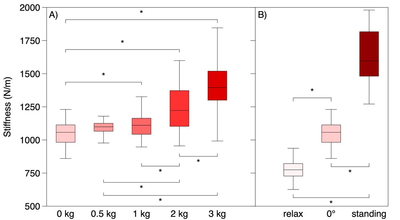
Changes in AT stiffness between the different conditions were calculated using Friedman test for related samples. Significant differences (p<0.01) were found between all contraction levels except for 0–0.5 kg (p=0.9); 0.5–1 kg (p=0.9); 1–2 kg (p=0.7) and between the different positions (figure 2).

Construct validity. Box plot showing the stiffness median and IQR values of the entire sample for the different contraction levels (A) and for the different positions (B). *p<0.01; statistical significant difference between the different conditions.
Discussion
The present study evaluated the absolute and relative reliability of the myotonometry to measure the stiffness of AT in different conditions. Even though the MyotonPRO was developed to evaluate the stiffness and tone of skeletal muscle, the present study shows the transferability of this technology and that measuring stiffness of tendon tissue is feasible and reliable. Intra-rater and inter-rater reliability ranged from high to very high in most of the tested conditions. Moreover, stiffness changes between the different contraction intensities and the different positions was detected, demonstrating the construct validity of this device.
One of the conditions in the current study was assessing AT stiffness with the person in a relaxed position with the feet hanging freely over the edge of the examination bed. A previous study25 also showed high intra-rater reliability when the AT was evaluated in a relaxed position (ICC: 0.80 in healthy participants). A further study evaluating the effect of dorsiflexion of the ankle joint on tendon stiffness18 showed high to very high intra-reliability and inter-reliability when measured in a relaxed position (ICC >0.88). Our results strengthen these previous observations by confirming high to very high intra and inter-rater reliability (ICC >0.86), now demonstrated in a larger sample. The stiffness values vary slightly between studies with an average of 873 N/m reported by Finnamore et al,25 776.11±71.70 reported by Liu et al,18 and 771±105.3 N/m observed in the present study. Nevertheless, collectively the current and previous findings confirm that MyotonPRO is a reliable device to evaluate AT stiffness in a relaxed position on the same day, between different days and between different operators.
Stiffness values increased from 1043±132.3 N/m when tendon was assessed in a 0° ankle dorsiflexion position with 0 kg of contraction to 1381±214 N/m for the isometric contraction at 3 kg. The ICC values showed high to very high intra-rater and inter-rater reliability for the majority of the isometric contraction levels (see table 2). Moderate intra-rater and inter-rater reliability was found for the 0.5 kg contraction level and the level of reliability did not exceed the cut-off point of ICC =0.6 considered as the threshold for clinical relevance.34 The low reliability for the contraction at 0.5 kg is likely due to the difficulty in maintaining the force feedback stable during such a low contraction of the plantar flexors. A previous study18 evaluating AT stiffness at different ankle angles showed similar results to the present study with an average stiffness of 1143 N/m at 0° of dorsiflexion compared with 1043±132 N/m found in the current study. Liu et al18 did not examine reliability at different ankle angles, however, the data reported in this article demonstrated a statistically significant increase in tendon stiffness due to the change in ankle dorsiflexion angle from 1143 N/m at 0° to 1329 N/m at 30°. These results are in line with the present study, which showed a significant increase in tendon stiffness with contractions of increasing intensity. Thus, the MyotonPRO can reliably detect changes in AT stiffness whether the tendon is stretched from contraction of the triceps surae muscle or stretched as a result of a change in the angle of the ankle.
Another study20 analysing the tissue properties of the AT and plantar fascia in healthy volunteers found slightly different stiffness values when the tendon was measured at 0° of dorsiflexion. The article reports a mean (±SD) of 848 (±95.73) N/m and 847.3 (±101.5) N/m for the left and right AT, respectively, whereas we observed higher values with an average stiffness of 1043±132 N/m.
Tendon structural and mechanical adaptations are strongly related to the load magnitude exerted to the musculotendinous complex3 5 and the majority of interventions targeting pathological tendon tissue are loading-based exercise regimens.36–40 The current results support the evaluation of the tendon tissue behaviour during load with a reliable and easy to use device could be helpful to detect possible mechanical adaptation following different rehabilitation protocol.
Stiffness of the AT was also evaluated in standing position in this study to verify the feasibility of MyotonPRO to analyse the tendon in a weight-bearing position. Intra-rater reliability was very high and inter-session reliability was high. Conversely inter-rater reliability in the standing position was moderate. It is important to note, however, that in the standing position the average stiffness values were 1596 (±339) N/m which is approaching the upper limit that the device is able to detect. The lower reliability could be a consequence of the measurement threshold of the device such that when it is close to the higher limit, there were difficulties in providing a mechanical impulse strong enough to give a proper oscillation wave. Morgan et al19 found similar results in a population of healthy participants in a weight-bearing position. Despite the possible technical limitation of analysing tendon stiffness in a weight-bearing position, the results demonstrate good intra-rater reliability but only moderate inter-rater reliability.
This is the first study to evaluate changes in AT stiffness between different contraction levels using the MyotonPRO. The results confirm a significant difference in tendon stiffness between the different contraction levels. Although stiffness changes can be detected across different contractions, the levels with similar intensity (0–0.5 kg (p=0.9); 0.5–1 kg (p=0.9); 1–2 kg (p=0.7)) showed similar stiffness values likely as the difference between consecutive contraction levels was not sufficient to change the mechanical properties of the tendon. These results are consistent with those reported in a previous sonoelastography evaluation41 showing an increase of tendon elastography colour scale index when the AT was gradually put under tension by isometric contractions of increasing intensity. In further support of the construct validity, pairwise comparisons showed a difference in AT stiffness between the different positions (relaxed, 0° ankle angle and standing). Similarly, a previous study using shear wave sonoelastography42 showed an increase in wave velocity, indicating a hardening of the AT, when the ankle angle changed from a plantarflexed position to 0° of dorsiflexion.
There are some limitations of this study which should be noted. Healthy participants were recruited and stiffness was evaluated in only one point of the tendon and it is therefore unknown whether variation may exist in different regions of the tendon. Another potential limitation was the selection of the contraction intensities. The upper limit of this ramp was chosen because after a certain amount of load (approximately 5 kg) the device cannot detect stiffness values. The small changes between the different contraction levels could be not sufficient to fully describe the behaviour of the tendon during load.
Conclusions
MyotonPRO is a reliable tool to measure AT stiffness in different conditions including when the tendon is in a relaxed position and under load. Reliability is confirmed for the same rater, between different days and between two raters. Construct validity was supported by changes of tendon stiffness during the explored contractions and ankle positions. MyotonPRO can be implemented, as a ready to use device, in the evaluation of tendon tissue mechanical properties.
Acknowledgments
We thank the students involved in the present study at the University of Applied Sciences and Arts of Southern Switzerland (SUPSI). We especially thank the Thim van der Laan Foundation, Landquart, Switzerland for financial support.
References
Footnotes
Contributors AS, DF, RC and MB designed and planned the study. AS and MB collected the data. All authors were involved in the drafting and revision of the manuscript and approved the final version of the article.
Funding The authors have not declared a specific grant for this research from any funding agency in the public, commercial or not-for-profit sectors.
Competing interests None declared.
Patient consent for publication Not required.
Ethics approval The study was approved by the Ethics Committee of Canton Ticino (ID:2018–01483/CE3391) and written informed consent was provided by the participants.
Provenance and peer review Not commissioned; externally peer reviewed.
Data availability statement Data are available on reasonable request.