Article Text

Abstract
Objective To investigate the influence of trunk and lower limb motion on electromyography (EMG) muscle activity and recruitment patterns around the shoulder.
Design Systematic review.
Data sources MEDLINE, CINAHL, PEDro, AMED, PubMed, Cochrane Central Register of Controlled trials, Cochrane Database of Systematic reviews, SportsDiscuss and PROSPERO.
Eligibility criteria Studies investigating both multiregional kinetic chain (KC) shoulder exercises and localised non-kinetic chain (nKC) shoulder exercises in healthy subjects under the same experimental conditions were included in this review.
Results KC exercises produced greater EMG activation levels in 5 of 11 studies for the lower trapezius. Of the remaining studies, five found no difference between the exercise types and one favoured nKC exercises. KC exercises produced greater EMG activation levels in 5 of 11 studies for the serratus anterior. Of the remaining studies, three reported the opposite and three found no significant difference between the exercise types. nKC exercises produced greater EMG activation in infraspinatus in three of four studies. KC exercises produced the lowest trapezius muscle ratios in all studies. Studies investigating the upper trapezius, middle trapezius, supraspinatus, subscapularis, biceps brachii, latifissimus dorsi, pectoralis major, deltoid, and trapezius and serratus anterior ratios showed inconsistency.
Conclusion This review found evidence that integrating the KC during shoulder rehabilitation may increase axioscapular muscle recruitment, produce lower trapezius muscle ratios and reduce the demands on the rotator cuff. Stepping appears preferable to squatting.
PROSPERO registration number CRD42015032557, 2015.
- shoulder
- rehabilitation
- exercise rehabilitation
- evidence based review
- sports rehabilitation programs
This is an open access article distributed in accordance with the Creative Commons Attribution Non Commercial (CC BY-NC 4.0) license, which permits others to distribute, remix, adapt, build upon this work non-commercially, and license their derivative works on different terms, provided the original work is properly cited, appropriate credit is given, any changes made indicated, and the use is non-commercial. See: http://creativecommons.org/licenses/by-nc/4.0/.
Statistics from Altmetric.com
What is already known?
Kinetic chain (KC) exercises are often advocated over an isolated local shoulder exercise approach during the rehabilitation of the shoulder, but its relevance is not well understood.
This review suggests that integrating the KC into shoulder rehabilitation exercises may enhance axioscapular muscle recruitment, produce lower trapezius muscle ratios and reduce the demands on the rotator cuff when compared with non-kinetic chain (nKC) exercises.
What are the new findings?
How the KC is integrated is key: stepping appears preferable to squatting.
KC exercises using lower quadrant weight transference may reduce the demands on the rotator cuff. nKC exercises may be preferable when the rehabilitation goal is to isolate and strengthen the rotator cuff.
Introduction
During sporting and non-sporting activities, the shoulder complex works as an integral part of the whole musculoskeletal system and not in isolation.1–6 The term kinetic chain (KC) refers to the sequential task specific activation of body segments during functional movement patterns.1 3 An efficient KC will generate, summate and permit efficient mechanical energy transfer throughout the whole chain contributing to function.1 4 Inefficiency within the KC at any ‘link’ has the potential to detrimentally affect force transfer to adjacent segments,1 3 4 which may require other components of the chain to increase their contribution to accommodate the energy loss.1 4 This has been postulated as a predisposing factor that increases the risk of shoulder injury and pain in overhead athletes.1 3 7–9
In tennis, the leg and the trunk generate 50%–55% of the total kinetic energy required for the serve.3 4 Lumbopelvic–hip stability and gluteal muscle activation are essential requirements for an efficient baseball pitch.10–14 Reduced hip abduction strength and hip range of motion have also been associated with increased risk of shoulder and elbow injury in throwing athletes.1 7–9 15–17 In addition, substantially higher rates of energy transfer through the shoulder complex have been observed during a tennis serve in injured players compared with their uninjured counterparts.4 Overhead athletes with lower limb injuries also appear to have an increased risk of upper limb injuries and pain,1 4 7–9 15–19 as well as reduced performance.4 20–22 Lower limb peak power has been found to be the primary determinant of throwing velocity in elite handball players21 and also strongly correlated with sprint-swim speed in freestyle swimmers.23 In addition, maximum velocity and muscle mass of the lower limb have been reported to be significantly correlated with javelin throwing performance in elite athletes.20
Isolated shoulder exercises may not address the altered global muscle activation patterns observed in those with shoulder injuries.1–3 6 Isolated shoulder exercises may address local strength deficits, yet local strength deficits are not definitively associated with shoulder injury.24–30 Consequently, clinicians frequently advocate the inclusion of lower extremity and trunk motion into shoulder rehabilitation programmes to optimise efficient energy transfer throughout the whole KC.1–3 5 6 31
The relevance of a KC approach over an isolated local shoulder exercise approach during the rehabilitation of the shoulder is not well understood. Therefore, the primary aim of this study was to investigate the influence of trunk and lower limb activity on the electromyography (EMG) and muscle recruitment patterns around the shoulder in a non-symptomatic population. A secondary aim was to identify deficits in current knowledge that would provide guidance for future research to better understand the relevance of the KC in normal function and rehabilitation.
Methods
The principles of the participants, interventions, comparisons, outcomes and study design (PICOS) criteria were followed and conducted in accordance with the Preferred Reporting Items for Systematic Reviews and Meta-Analyses guidelines.32
Eligibility criteria
Studies
Preliminary searching identified the need to include non-randomised studies in the review to ensure that all studies relevant to the research question were evaluated. Studies considered level 3 and above on the 2011 Oxford Centre for Evidence-Based Medicine (OCEBM)33 were eligible for inclusion. Only studies published in English were evaluated, and no publication date or status restrictions were imposed. Only studies interpreting original data with ethical approval were included.
Participants
Studies conducted on healthy adults (or with a healthy subject cohort group) over 16 years of age were included. Studies investigating wheelchair users were excluded as the aims were to assess the role of leg and trunk motion on shoulder function.
Intervention
Studies investigating the use of both KC shoulder exercises and local non-kinetic chain (nKC) shoulder exercises under the same experimental conditions were included. KC exercises investigated in studies were not required to be exclusively direct movement comparisons of their nKC counterparts. Additionally, both open KC and closed KC were included, assuming they also involved activating the muscles of the trunk or the lower limb and/or the motion of these segments.
Outcomes
Studies evaluating EMG activity using both surface and indwelling electrodes were included.
Search strategy
Studies were identified by searching the following databases: MEDLINE (in-process and other non-indexed citations and OVID MEDLINE), CINAHL, PEDro, AMED, PubMed, Cochrane Central Register of Controlled trials, Cochrane Database of Systematic reviews, SportsDiscuss and PROSPERO. Searches were conducted on 10 May 2018 and reran on 7 September 2019.
Abstract and subject heading search phrases were developed and finalised jointly by three reviewers (ER, CM and GY) following extensive background research (table 1).
PICOS abstract and subject heading search phrases
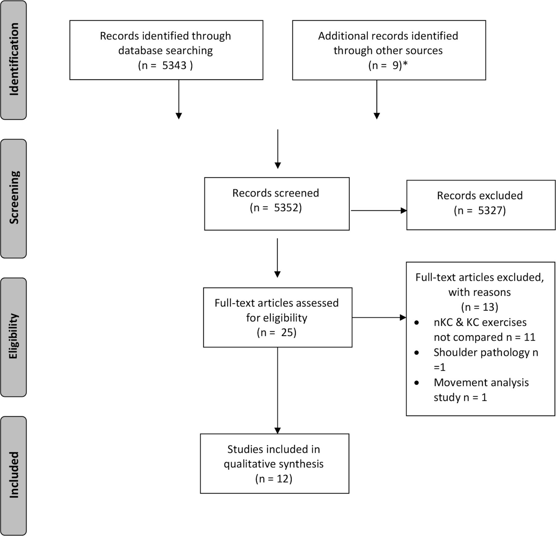
The outcome of interest in this study was EMG, and to ensure all relevant studies were considered, such as those where EMG did not appear in the title or abstract but was used as an outcome measure, it was necessary to broaden the terms used. Similarly, while the intervention of interest in this study was the KC, to ensure the inclusion of all relevant studies, broader search terms in relation to exercise therapy were used (table 1). Titles and abstracts were independently reviewed for eligibility, and any discrepancies were discussed. Disagreements on inclusion of articles were resolved through discussion between ER and CM and, when necessary, a third reviewer (JG). Only studies meeting the full eligibility criteria were considered for review. Manual searching of the reference lists of included articles to identify any additional studies meeting the inclusion criteria was also conducted (figure 1).

Preferred Reporting Items for Systematic Reviews and Meta-Analyses flow diagram24 illustrating the systematic process of inclusion and exclusion criteria application, which generated the final number for analysis in this systematic review. *Manual searching through reference lists/bibliographies, consultation with JG and discussion with other clinical experts regarding unpublished research. KC, kinetic chain; nKC, non-kinetic chain.
Methodological quality assessment
Methodological quality of included studies was independently assessed by two reviewers (ER and CM) using the modified Downs and Black tool (mD&B).34–41 The tool constitutes a 27-point checklist split across the following subsections: reporting, external validity, internal validity (bias) and internal validity (confounding). The total score for this tool ranges from 0 to 28, with a higher score indicating higher methodological quality.35 39 40 Disagreement among the reviewers was resolved by consensus. Each paper was assigned a quality grade of ‘excellent’ (24–28 points), ‘good’ (19–23 points), ‘fair’ (14–18 points) or ‘poor’ (<14 points), as documented previously.42
Risk of bias (ROB)
O’Connor et al42 have challenged the use of the mD&B tool, suggesting it may fail to identify ROB in all studies. Therefore, specific ROB assessment was also undertaken through the application of a dedicated ROB tool: ROBINS-1.43 Two reviewers (ER and CM) applied this tool independently with 100% agreement.
Electromyography
EMG has been suggested to be the most reliable tool researchers have to evaluate the complex activation patterns of muscles during movement.44 However, cross talk may influence the validity of EMG through the contamination of signals.45–48 Surface electrodes are more susceptible to cross talk contamination than indwelling electrodes.45 Recent evidence indicates that surface electrode recordings overestimate infraspinatus and latissimus dorsi activity due to pick up from surrounding muscles.45 49 Surface electrodes are also susceptible to geometric displacement. When placed on the area overlying the inferior slips of serratus anterior it has been shown that activity levels in this muscle during shoulder movements are underestimated compared with that recorded using indwelling electrodes due to this type of displacement.50
In light of these known limitations, a scoring tool to evaluate EMG methodological quality was developed for this study (online supplementary file 1).
Supplemental material
The evaluation tool was applied to five articles by ER, and the results were discussed with two other reviewers (KG and MH) with extensive EMG experience until there was full agreement. The evaluation tool was then applied to the remaining seven articles by ER.
EMG methodological quality scoring tool
The EMG methodological quality scoring tool was developed based on the Standards for Reporting EMG data as endorsed by the International Society of Electrophysiology and Kinesiology.51 It was divided into six domains with a maximum number of points available in each: EMG detection of signal (five points), processing signal (two points), EMG levels/patterns (two points), EMG timing (one point), procedures (two points) and analysis (one point). A percentage quality score was generated for each study based on the total number of points earned divided by the total number of points available. All domains were scored using a simple ‘yes’ equals one point and ‘no’ equals zero points, with the exception of the first domain, EMG detection of signal. The first subsection in this domain relates to electrode type and asks, ‘Was the selection of electrode (surface or indwelling) appropriate?’ If all the electrode types used were appropriate for each muscle under investigation, then yes would be ticked and a score of 1 awarded. However, for studies in which both yes and no answers were recorded for different muscle groups, a score between 0 and 1 was awarded based on the total number of muscle groups listed under yes divided by the total number of muscle groups listed under both yes and no columns. For example, if three of four muscle groups investigated scored yes, then a score of 0.75 would be awarded for this section and added to the remainder of the points earned in this domain.
For ease of between-study ranking only, overall percentages were awarded to each study.
Data analysis
Data relating to the research question was extracted by ER and CM and inputted into a Microsoft Excel V.16.10 spreadsheet. Data extraction was piloted by ER and CM, and any misunderstandings and disagreements were addressed through consultation. A third reviewer (GY) was available to arbitrate any unresolved issues but was not required. Of the 12 studies included in the review, disagreement between the reviewers (ER and CM) with data extracted occurred in one study.52 This was resolved by contacting the author to clarify EMG amplitudes to ensure accurate data extraction.
Due to the small sample sizes and the heterogeneity of the outcomes assessed, a meta-analysis or statistical assessment of the outcomes was not performed, and a narrative analysis was undertaken.
Results
Twelve articles met the inclusion criteria for this review (online supplementary file 2).52–63 Seventeen muscle groups were investigated in 204 participants without symptoms.
Supplemental material
Study characteristics
Studies were assessed to be either level 2 or 3 OCEBM evidence.33 All EMG results were expressed as a percentage of maximal voluntary contraction (MVC). Two studies also provided additional information for axioscapular muscle ratios.57 63 All studies used surface EMG with two also using fine wire intramuscular electrodes in supraspinatus and infraspinatus.60 62 Smith et al60 also investigated the upper subscapularis. A total of 85 exercises were investigated: 45 individual KC exercises and 40 nKC exercises.
The muscles investigated in each study varied, with lower trapezius (n=11), serratus anterior (n=11) and upper trapezius (n=10) comprising the muscles most commonly evaluated. Less commonly evaluated were infraspinatus (n=4), supraspinatus (n=2) and latissimus dorsi (n=2), with the remaining muscle groups investigated only in individual studies.
Methodological quality
Ten out of the 12 studies were rated ‘fair’,52–59 61 62 with the remaining 2 rated ‘poor’60 63 on the mD&B (table 2).
Downs and black methodological quality assessment results
The ROB was scored as ‘low’ across all studies, with only one domain, bias due to confounding, showing a moderate ROB (online supplementary file 3).
Supplemental material
With respect to EMG methodological quality, 7 of the 12 studies were awarded greater than 80%,52 53 55 57–59 63 two between 70% and 79%56 61 and three studies less than 70%54 60 62 (table 3), with a mean percentage score of 78% (SD=10%).
EMG methodological quality evaluation
Comparative EMG activity between KC and nKC exercises
Axioscapular muscles
The results suggest that KC exercises may preferentially activate and produce higher EMG amplitudes in the whole trapezius and lower trapezius when compared with their nKC counterparts. Conflicting results exist in relation to upper trapezius and middle trapezius (online supplementary file 2 and table 4). Removal of studies with the lowest methodological quality for both the mD&B and EMG methodological quality assessment did not add clarity to these conflicting results.
To show whether studies favoured nKC or KC exercise for overall EMG activation levels
Trapezius
Of the studies investigating the upper trapezius (n=10),52–54 56–60 62 63 four found no significant difference in activation levels between KC and nKC exercises52 53 58 60; four found KC exercises elicited greater EMG activation than nKC exercises54 56 59 62; one study63 found nKC exercises elicited greater EMG activation than KC exercises; and one study did not provide a direct KC versus nKC comparison.57 Of the studies investigating the middle trapezius (n=4),57 58 60 63 one showed no significant difference in activation levels between KC and nKC exercises60; one did not provide enough comparative data to determine the presence of any significant difference57; and two studies provided conflicting results.58 63 Of the 11 studies investigating the lower trapezius,52–54 56–63 5 demonstrated consistently higher EMG activation levels during KC exercises compared with nKC exercises.56–58 62 63 Five studies showed no difference,52–54 59 60 and one study found nKC exercises elicited greater EMG activation than KC exercises.61 One study investigated the whole trapezius53 and found the KC exercise ‘high scapula retraction in static unipedal squat’ elicited higher EMG activation than nKC ‘high scapula retraction in sitting’ (online supplementary file 2). Removal of studies with the lowest methodological quality score for both the mD&B and EMG methodological quality assessment saw seven studies remaining. Of these, three favoured KC exercises in eliciting the highest EMG activation levels56–58; one favoured nKC exercises61; and three reported no significant difference between the two.52 53 59
Serratus anterior
Of the studies investigating the serratus anterior (n=11),52 54–63 five reported KC exercises produced significantly higher EMG activation levels compared with nKC exercises55 57–60; three studies reported the opposite52 61 63; two studies found no significant differences54 56; and one study found an equal total number of KC exercises produced greater EMG activity than nKC exercises and vice versa (online supplementary file 2 and table 4).62 Removal of studies with the lowest methodological quality score for both the mD&B and EMG methodological quality assessment saw seven studies remaining. Of these, four favoured KC exercises for eliciting the highest EMG activation levels55 57–59; two favoured nKC exercises52 61; and one found no difference between the two.56
Rotator cuff
Non-KC exercises produced higher EMG amplitudes in the infraspinatus when compared with their KC counterparts in three of four studies,52 61 62 with the remainder showing no significant different between exercise types.60 One study investigating the upper subscapularis62 reported no significant difference in EMG activation levels between the two exercise types. Two studies investigated the supraspinatus60 62 and found no significant difference in EMG amplitudes between KC and nKC exercises60 and an equal total number of KC exercises producing greater EMG activity than nKC exercises and vice versa (online supplementary file 2 and table 4).62 Removal of studies with the lowest methodological quality score for both the mD&B and EMG methodological quality assessment saw two studies remaining, both favouring nKC in eliciting the highest EMG activation levels.52 61
Glenohumeral joint prime movers
The results from this review show an unclear picture in relation to whether KC or nKC exercises preferentially activate muscles considered prime movers of the glenohumeral joint. No significant differences in EMG activity was found for biceps brachii60 latissimus dorsi,54 55 pectoralis major59 or the middle deltoid.60
Of the studies investigating the anterior deltoid (n=5),52 56 59 60 62 an equal total number of KC exercises produced greater EMG activity than nKC exercises and vice versa (online supplementary file 2 and table 4). Removal of studies with the lowest methodological quality score for both the mD&B and EMG methodological quality assessments saw three studies remaining, two of which favoured nKC exercises for eliciting highest EMG activation levels52 56 and one favouring KC exercises.59
Trunk, pelvis and lower limb
One study investigated the external oblique, ipsilateral gluteus maximus, contralateral gluteus maximus and the femoral adductor muscles55 and found KC exercises produced significantly greater EMG activity in each muscle group compared with that elicited during nKC exercises (online supplementary file 2 and table 4). This study was of fair methodological quality on the mD&B and scored in the highest category (greater than 80%) on our EMG methodological quality assessment.
Muscle ratio comparisons
Upper trapezius:lower trapezius ratio (UT:LT)
KC exercises produced lower UT:LT ratios when compared with their nKC counterparts in one study.63 Ratios were less than 1 for all exercises except nKC standing scaption. KC standing external rotation plus trunk and hip rotation produced the lowest ratio (0.2) (table 5).
To show whether studies favoured nKC or KC exercises for axioscapula muscle ratios
Maenhout et al57 did not provide information regarding a direct KC versus nKC ratio comparison. KC exercise ‘kneeling push-up plus (KPP) with heterolateral leg extension’ produced the lowest ratio (1.03).
Upper trapezius:serratus anterior ratio
Maenhout et al57 reported varied ratios across all exercises but found nKC exercise ‘KPP’ to be significantly lower than KC exercise ‘KPP with heterolateral leg extension’ and ‘KPP with heterolateral leg extension on a wobble board’. KC exercise ‘KPP with homolateral leg extension’ produced the lowest overall ratio (0.4).
Yamauchi et al63 found KC exercises produced significantly lower ratios than nKC exercises in three out of five comparisons, with nKC exercises producing significantly lower ratios in the remaining two. Non-KC exercise ‘standing external rotation at 90 degrees abduction’ produced the lowest ratio (0.5) (table 5).
Upper trapezius:middle trapezius ratio (UT:MT)
Yamauchi et al63 found KC exercises produced lower UT:MT ratios when compared with their nKC counterparts. The mean ratio for all exercises regardless of type or category was 1.15 (SD=0.9), with KC exercise ‘standing external rotation plus hip and trunk rotation’ producing the lowest ratio (0.3). During direct KC and nKC comparisons, only KC exercise ‘prone scapula retraction at 90 degrees abduction plus trunk rotation’ (0.6) and nKC ‘prone scapular retraction at 90 degrees abduction’ (0.9) recorded a ratio of less than 1 (table 5).
Maenhout et al57 did not provide information regarding a direct KC versus nKC comparison. However, the mean ratio for all exercises regardless of type or category was 1.54 (SD=1.5), with KC exercise ‘KPP with heterolateral leg extension on a wobble board’ producing the lowest ratio (1.44).
Yamauchi et al’s63 study scored poor on the mD&B methodological quality assessment but scored in the highest category (greater than 80%) on our EMG methodological quality assessment.
Discussion
This review aimed to investigate whether the addition of lower limb and trunk motion into shoulder exercises influenced EMG activity and recruitment patterns around the shoulder complex. Overall, the findings suggest that integrating the KC into shoulder exercises may enhance axioscapular muscle recruitment, produce lower trapezius muscle ratios and reduce the demands on the rotator cuff.
KC and the axioscapular muscles
Lower trapezius
A preferential effect favouring motion in the form of lateral weight transference as a KC integration strategy compared with movements such as bipedal squatting was observed in this review. Of the five studies56–58 61–63 that favoured KC exercises, 4 involved significant weight transference through the lower limb57 58 61–63 and 1 combined hip and knee extension with trunk and thoracic rotation.58 The results in the remaining six studies suggest that sequential lateral weight transference may be key to eliciting higher lower trapezius activation levels.
Tsuruke & Ellenbecker61 elicited higher activation levels in the lower trapezius during nKC quadruped shoulder flexion compared with the ‘lawn-mower’. Closer inspection of the techniques employed during these two commonly prescribed KC exercises reveals a disparity between exercise execution and the recommendations within the literature.1–5 In Tsuruke & Ellenbecker,61 the ‘lawn-mower’ appears to have been without any lateral weight transference through the lower limb.1–5 However, when evaluating this comparison in isolation it is not possible to infer whether this difference relates to suboptimal KC sequencing or the inherent differences in gross movement patterns between each exercise.
A direct movement comparison between lateral rotation of the shoulder in standing with and without the addition of trunk rotation, was assessed by Yamauchi et al.63 The end position during this latter exercise is similar to the end position of the ‘lawn-mower’ described by Tsuruke & Ellenbecker.61 In Yamauchi et al’s.63 study the arm was supported with a towel, the head is observed to follow the movement and although the feet remain facing forwards, weight transfer towards the ipsilateral leg can be observed. This method of sequencing is arguably more in keeping with the frameworks that exist in the literature1–5 and suggests that the order in which the KC is integrated may affect lower trapezius activation levels. However, it is not clear whether contributions from a particular KC component lead to the higher lower trapezius activation levels observed.
The components of KC sequencing may be evaluated further if the findings from Tsuruike et al61 and Yamauchi et al63 are combined with the findings of Nagai et al.59 In Nagai et al,59 no significant difference was found in lower trapezius activation levels between bilateral shoulder flexion in sitting with or without the addition of trunk rotation. Unlike Yamauchi et al,63 the lower quadrant was omitted from this sequencing pattern. Exercise instructions also differed with Yamauchi et al63 requesting subjects maximally rotate the trunk and hips during shoulder rotation and Nagai et al59 instructing a more segmented approach of rotation followed by shoulder flexion. These findings reinforce the potential importance of fluid proximal-to-distal lateral weight transference through the lower quadrant during KC integration.
The preferential effects of lateral weight transference as a KC integration strategy on lower trapezius is also observed in studies where differences were reported but not deemed statistically significant.52–54 60 Studies that employed a KC exercise variant that involved stepping all yielded higher activation levels in the lower trapezius than their nKC counterparts.54 60 Conversely, studies employing a static bipedal stance without dynamic lateral weight transference did not yield higher lower trapezius activation levels than their nKC counterpart.52 53 59 One study60 investigated the effect of an attempted overhead ‘reach’ in bipedal stance and during a stepping motion as well as an attempted cross body ‘reach’ in bipedal stance and during a stepping motion. Arm immobilisation removed glenohumeral joint motion as a variable rendering the ‘reach’ a shoulder girdle shrugging motion. The addition of stepping revealed a trend of higher lower trapezius activation levels in all exercises, although this was not deemed statistically significant. Although clinical and statistical significance do not always equate, no data exist to report whether this observed trend was clinically relevant.
When this narrative analysis is examined in conjunction with the results from the methodology quality assessments, more studies (n=3) favour KC exercises56–58 over nKC exercises61 (n=1) for eliciting highest EMG activation level in the lower trapezius. Of the three studies that did not show consistent differences between either KC and nKC exercises, the KC exercises investigated all involved static bipedal postures and did not involve any fluid lateral weight transference through the lower quadrant during EMG data collection.
Serratus anterior
Unless surface electrodes are placed at the angle of testing during isometric contractions, they weakly correlate with intramuscular electrodes from 90 degrees of elevation upwards during activities where serratus anterior would be expected to be active.50 It is notable that studies in this review favouring nKC exercises over KC exercises, or finding no significant difference between the two, investigated exercises from 90 degrees elevation upwards using surface electrodes.54 56 61
One study63 found nKC exercises produced higher serratus anterior activation levels than KC exercises in 2 out of 3 comparisons. Standing external rotation at 90 degrees abduction and prone retraction at 145 degrees abduction were evaluated with and without the addition of trunk rotation. Given the potential for geometric electrode displacement during trunk rotation, coupled with Hackett’s et al’s.50 findings, the validity of this comparison is challenged. Furthermore, the comparison which favoured KC inclusion involved lateral rotation with the arm in neutral with and without thoracic rotation.63 With the arm below 90 degrees and serratus anterior activity expected to be low during both exercises, this comparison is arguably more valid.50
Of the studies that favoured KC exercises,55 57 58 60 all involved significant weight transference or unilateral loading through the lower quadrant. As geometric displacement was likely in 3 of these four studies,55 57 58 it could be argued that the differences noted in favour of KC exercises were actually under-estimated.50 The results from Smith et al’s.60 study, where the addition of stepping with the arm immobilised increased serratus anterior activation to the highest level seen across all 12 studies (199% MVC) when compared with bipedal stance, further supports the notion that KC integration enhances serratus anterior activation levels.
KC and axioscapular muscle ratios
Restoration of balanced co-activation of the axioscapular muscles is considered an important component of shoulder rehabilitation programmes.64 A lack of activity in lower trapezius, middle trapezius and serratus anterior in combination with excessive upper trapezius activity has been described.57 64 As such, UT/LT, UT/MT and UT/SA ratios have been frequently evaluated in the literature with ratios of less than one being deemed desirable.57 63 64
One study in this review investigated trapezius muscle ratios across all exercises.63 KC exercises consistently produced lower UT/LT and MT/LT muscle ratios than their nKC counterparts. Standing external rotation of the shoulder at 0 degrees abduction with the addition of trunk and hip rotation produced the lowest UT/LT ratio (0.2) whereas prone shoulder retraction at 90 degrees abduction plus trunk rotation produced the lowest UT/MT ratios (0.6). This is partly in keeping with Cools et al64 who found favourable UT/MT and UT/LT muscle ratios during side lying external shoulder rotation at 0 degrees and prone horizontal shoulder abduction with external rotation at 90 degrees.
Cools et al64 concluded that eliminating the effect of gravity on upper trapezius lead to these favourable ratios. The findings of this review partly support this conclusion with the only two exercises to produce an UT/MT ratio of less than one being in the prone position. However, this inference does not explain why the most favourable UT/LT ratio in Yamauchi et al’s.63 study was noted during standing. Nor does it explain the significant decrease in UT/LT ratios noted when scaption through full range in standing (1.3) was compared with scaption with the addition of trunk and hip rotation (0.8). The addition of trunk and hip rotation, plus the observed lateral weight transfer that coupled this movement, appears to have enhanced recruitment of the lower trapezius.
Two studies investigated UT/SA ratios57 63 but only one directly compared ratios between KC and nKC exercises.63 Although Yamauchi et al63 found KC exercises produced lower ratios than nKC exercises in three out of five comparisons, two of the five favoured nKC exercise variants. During one of these comparisons, arm abduction angle was 90 degrees for both KC and nKC exercise variants and serratus anterior activation levels were expected to be high – criteria that render surface EMG most likely to under-report serratus anterior activation levels.50 The remaining comparison evaluated the addition of thoracic rotation to arm elevation at 145 degrees in prone. Where arm elevation angles were below 90 degrees and serratus anterior not expected to be overly active, KC exercises produced more favourable ratios.
The pitfalls of surface EMG as a valid outcome measure for serratus anterior might explain why UT/SA ratios of less than one were infrequent (table 5). Clinicians must therefore interpret any muscle ratio where surface electrodes were used for serratus anterior activity with caution. The limitations of surface EMG as a valid outcome measure for serratus anterior may also explain why Cools et al64 were unable to find any exercise for optimising UT/SA ratios.
KC and the rotator cuff
Findings from this review suggest that reduced contribution from the KC has the potential to influence force transfer to distal segments,1–4 with nKC exercises preferentially activating the infraspinatus and supraspinatus.52 60–62 However, only four studies investigated the infraspinatus with three of these considered to be of poor methodological quality.52 60 61 In addition to this, two studies used surface EMG during movements where infraspinatus would be expected to be both active and inactive at different phases of the movement,52 61 a scenario where intramuscular electrodes are required to give valid infraspinatus measures.45 If this is taken into consideration, only two studies that investigated both infraspinatus and supraspinatus remain,60 62 one of which found no difference between KC and nKC exercises.60 Furthermore, overall activity in the remaining study was consistently low for infraspinatus (2%–14% MVC) and low to moderate to supraspinatus (4%–29% MVC).65
Strengths & limitations of review
The strength of recommendations made by a systematic review relates directly to the quality of the studies evaluated.32 There was a moderate ROB due to confounding for all studies. However, the pitfalls of EMG as an objective measure render eliminating ROB for this domain unlikely.
Studies in this review were considered of ‘fair’ to ‘poor’ quality on the mD&B scale. However, it was considered that all domains in the mD&B tool were not equally pertinent to the quality of investigatory studies solely evaluating EMG amplitudes among healthy subjects. Consequently, items deemed most and least pertinent were highlighted as dark grey and light grey, respectively (table 2). If the items deemed least pertinent are excluded, this would theoretically increase all but one study65 to a quality grade rating of ‘good’ based on O’Connor et al’s.42 classification system. It is acknowledged, however, that this postulation adds a subjective element to an otherwise systematic process.
We acknowledge that no studies exist regarding the validity and reliability of our novel EMG methodology quality assessment method, and therefore caution must be taken when attaching meaning to these results. It is therefore recommended that readers use the percentages generated as a means of quality comparison between studies only until such time as this scoring method is evaluated further. In addition, as only studies written in English were included in this review, the authors are uncertain of the number or quality of any non-English language publications related to the research question. Consequently, a future systematic review that does not have this limitation may produce different results or conclude with different findings.
Not all studies included in this review investigated KC exercises where the KC segment involved provided a direct movement comparison of their nKC counterpart exercise. The authors acknowledge that in these circumstances, direct conclusions from the data cannot be drawn but instead use these comparisons to inform the narrative discussion around the interpretation of the collective results across all studies.
No metanalysis was performed in this review, which may be considered as a limitation. However, due to heterogeneity among studies in relation to the exercises undertaken, the electrode type, placement and protocol followed, as well as the variations of ROB among the included studies a metanalysis was not performed, as recommended in the Cochrane guidelines.37
Conclusion
This review found evidence that may suggest integrating the KC into shoulder rehabilitation exercises may enhance axioscapular muscle recruitment, produce lower trapezius muscle ratios and reduce the demands on the rotator cuff. The manner in which the KC is integrated may be key in eliciting these potentially clinically favourable outcomes, with consistent evidence favouring lateral lower quadrant weight transference methods, such as stepping, over common KC integration strategies such as squatting. Conflicting evidence suggests that nKC exercises are preferable when the rehabilitation goal is to isolate and strengthen the rotator cuff, whereas KC exercises may be more suited when targeting enhanced efficiency. Caution is needed when the results of studies using surface EMG as an outcome measure are used to inform exercise selection, especially in relation to serratus anterior and ratios where the serratus anterior forms part of the formula. Further investigations are required to explore the role of lower quadrant weight transference in relation to optimising shoulder muscle recruitment patterns in both healthy and injured cohorts. Future research investigating the validity and reliability of our peer reviewed EMG methodological quality assessment tool is also warranted.
References
Footnotes
Twitter @Els_Richardson, @JeremyLewisPT, @Shouldergeek1, @ChrisMorgan10, @HalakiMark
Contributors All those involved in this systematic review met the criteria for coauthorship, and therefore no Contributorship statement was required.
Funding The authors have not declared a specific grant for this research from any funding agency in the public, commercial or not-for-profit sectors.
Competing interests None declared.
Patient and public involvement statement Patients were not involved in this research.
Patient consent for publication Not required.
Provenance and peer review Not commissioned; externally peer reviewed.
Data availability statement All data relevant to the study are included in the article or uploaded as supplementary information.