Article Text

Abstract
Background Soccer players are commonly affected by long-standing adductor-related groin pain (ARGP), but the clinical significance of MRI findings in these athletes is largely unknown. Our aims were (1) to evaluate whether MRI findings are associated with long-standing ARGP in soccer players, (2) to assess MRI findings in asymptomatic soccer players and non-soccer playing controls.
Methods This cross-sectional study included 28 male soccer players with long-standing ARGP, 17 male asymptomatic soccer players and 20 male asymptomatic non-soccer playing athletes of matching age and athletic exposure. Participants underwent identical standardised and reliable clinical examination, and MRI scans (3 T) of the pelvis performed by a blinded observer. Images were consensus rated by three blinded radiologists according to a standardised MRI evaluation protocol. The associations between clinical adductor-related findings and pathological MRI findings were investigated with χ2 statistics and OR.
Results Central disc protrusion (p=0.027) and higher grades of pubic bone marrow oedema (BMO; p=0.027) were significantly more present in symptomatic players than asymptomatic players. However, up to 71% of asymptomatic soccer players displayed different positive MRI findings, and asymptomatic soccer players had significantly higher odds (OR ranging from 6.3 to 13.3) for BMO, adductor tendinopathy and degenerative changes than non-soccer players.
Conclusions ARGP in soccer players was associated with central disc protrusion and higher grades of pubic BMO. Moreover, positive MRI findings were significantly more frequent in soccer players compared with non-soccer players irrespective of symptoms, suggesting that these MRI changes may be associated with soccer play itself rather than clinical symptoms.
- MRI
- Groin
- Soccer
- Lowever extremity
- Sporting injuries
Statistics from Altmetric.com
Introduction
Soccer is the most popular sport worldwide1 counting approximately 265 million professional and amateur players.2 The annual incidences of groin injuries are 12–16%,3 ,4 with adductor-related injuries being the most common type of groin injury,3 ,5 and the second most common type of muscle injury in soccer.6 Sprinting, kicking and change of direction7 ,8 induce considerable stress at the adductor musculotendinous complex, and seem to play an important role in relation to groin injury mechanisms in soccer.
MRI has been increasingly used to evaluate groin pain in athletes, because of its ability to visualise soft tissues.9 ,10 However, MRI studies are scarce, and comparing results between published papers is difficult mainly because of a lack of consensus regarding terminology and diagnostic terms, and a shortage of reliable assessments.11 Clinical examinations and MRI findings are often not reported in detail. To address these shortcomings, our research group has previously developed a standardised MRI evaluation protocol to be used for imaging of patients with sports-related long-standing hip and groin pain.11a The protocol is composed of specific predefined imaging characteristics indicative of the possible pathology seen around the symphyseal joint, the pubic bones and the adductor and rectus abdominis insertions at the pubic bones.
The clinical significance of positive MRI findings for adductor-related groin pain (ARGP) is currently uncertain.11 ,12 Moreover, athletic activity itself seems to be associated with radiographic degenerative changes at the symphysis13–15 and MRI-detected pubic bone marrow oedema (BMO),16–19 as well as with MRI changes in other asymptomatic joints.20–23 Therefore, we wished to compare soccer players with ARGP with asymptomatic controls according to our MRI evaluation protocol to investigate the relationship between clinical symptoms, soccer play and MRI findings.
The primary aim was to compare MRI findings between symptomatic and asymptomatic soccer players, and to assess whether positive MRI findings are associated with ARGP. The secondary aim was to compare asymptomatic soccer players and asymptomatic non-soccer playing athletes of matching age and athletic exposure to evaluate whether predefined MRI findings are more common in asymptomatic soccer players than in other asymptomatic athletes.
Material and methods
Study population
Data are based on a large cross-sectional cohort study investigating hip and/or groin pain, self-reported outcome, clinical data, muscle strength, range of motion and radiological findings in male soccer players.24 ,25 All participants provided written informed consent according to the Helsinki Declaration. The study was approved by the Danish National Committee on Health Research Ethics (H-2-2010-127) and the Danish Data Protection Agency (2011-41-5964).
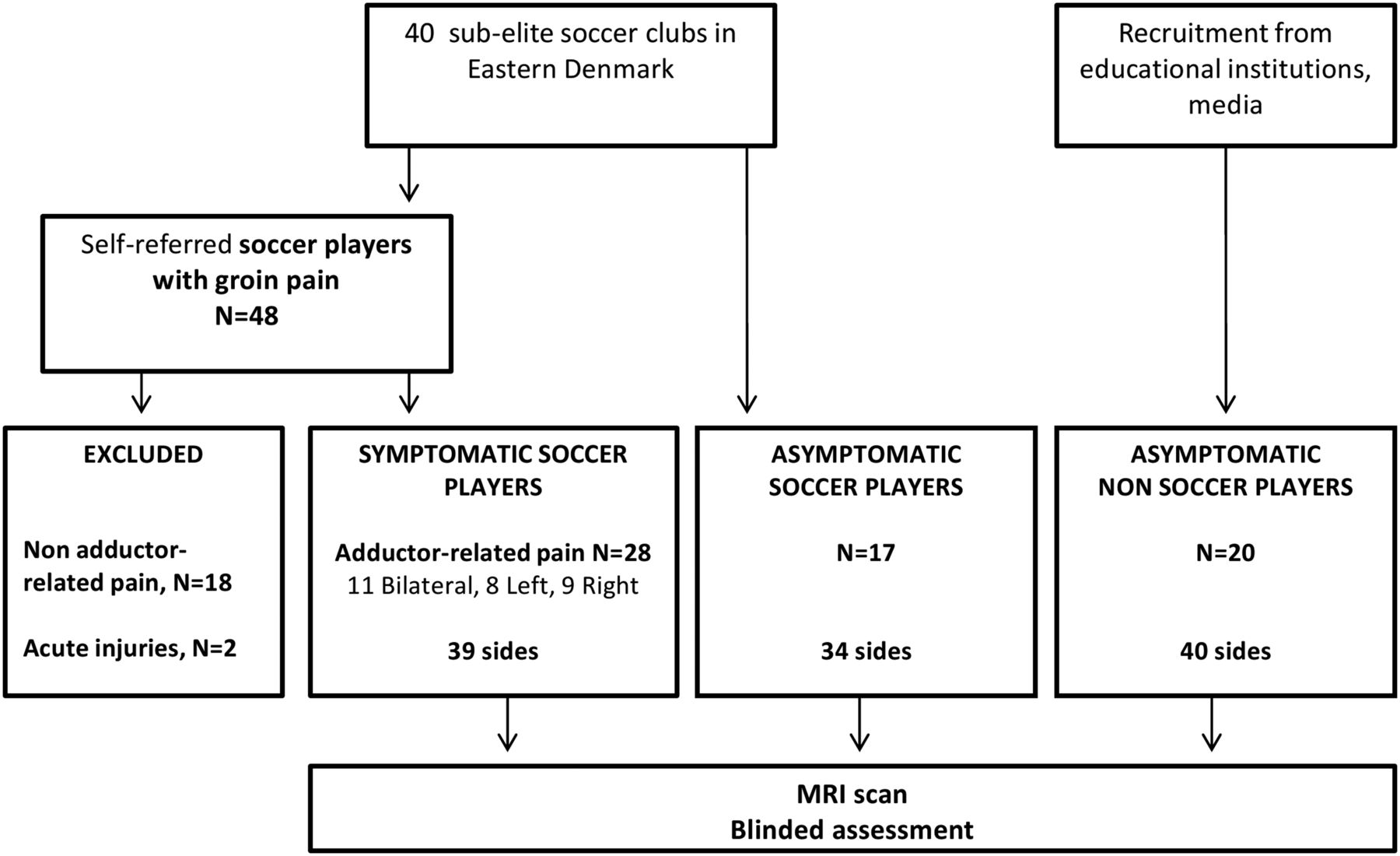
Forty sub-elite soccer clubs (Division 1–4) in Eastern Denmark were provided with oral and written information about the study in August 2011. Forty-eight male soccer players with hip and groin pain contacted us to participate. To include as many eligible injured players as possible in the symptomatic group, we decided to include players who had experienced ARGP for at least 4 weeks, without clinical signs of osteoarthritis, as 4 weeks is, in our experience, long enough to assume that pain is neither acute nor merely temporary.5 Twenty-eight players with ARGP were included, and 20 players were excluded: 18 did not have ARGP, and 2 had acute injuries (<4 weeks). Two control groups were also included. The first control group consisted of male soccer players, who were asymptomatic clinically and had no recollection of hip and groin pain experienced within the year before recruitment for this study, which had kept them out of training or games: 20 players were examined clinically, and three were excluded because they could not serve as asymptomatic controls (two had pain on palpation of the adductor tendons without having experienced pain when playing, and one experienced symptoms immediately prior to examination), leaving 17 asymptomatic players for inclusion.
The second control group consisted of 20 age-matched male non-soccer playing athletes to ensure that no significant difference in athletic activity level would be a confounder. These athletes had never played organised soccer, and they participated in different sports: fitness (N=9), martial arts (N=3), running (N=3), cycling (N=2), parkour (N=1), kayak (N=1) and basketball (N=1). Asymptomatic non-soccer players were recruited through advertising in sports clubs, educational institutions and fitness centres. They were given the contact details of a research assistant at our institution, who evaluated whether potential participants were eligible to be included in the study based on the personal information they provided. To ensure adequate blinding of examiners, our research assistant was not involved in performing clinical and radiological examinations, but organised schedules and bookings of all examinations. The blinded examiners (clinicians and radiologist) knew only the names and dates of birth of study participants, but had no access to clinical information. Moreover, study participants were instructed not to disclose to the clinicians and radiologists at the time of examination whether they were symptomatic, and whether they played soccer.
Thus, 65 male participants (age range 18–41 years; mean age 24.5±8 years) were included in this study. Age, weight, height, duration of pain, physical activity level and the Copenhagen Hip And Groin Outcome Score (HAGOS)26 were recorded for all participants. Only male participants were included to ensure that they were readily comparable because groin injuries seem to be more frequent in male than in female athletes,27 ,28 and because of anatomical differences in the groin region between sexes.29 The selection process is displayed in figure 1, and inclusion and exclusion criteria in table 1.
Inclusion and exclusion criteria for participation in the study

Selection of study participants.
MRI scanning protocol
MRI scans were acquired on a 3 T Siemens Magnetom Verio system (Siemens, Erlangen, Germany), participants lying supine with a surface coil (32-channel high-resolution body coil) centred at the pubic symphysis and covering the pelvic area. Eight sequences were performed, taking approximately 40 min, and are displayed with scan parameters in table 2. The axial oblique plane was tilted approximately 50° from the horizontal plane, and oriented parallel to the long axis of the superior pubic rami (figure 2), in order to enable visualisation of the adductor longus tendons along their long axes.30 All MRI scans were performed by a blinded radiologist (SB), unaware of the clinical information on study participants other than their name and date of birth, and stored in the institution's Picture Archiving and Communicating System (PACS).
MR scan parameters

Scan plane for axial oblique sequences, oriented parallel to the superior pubic rami on a sagittal short tau inversion recovery (STIR) image.
Clinical examination
All participants underwent a standardised and reliable clinical examination31 to identify specific clinical entities using the approach described by Hölmich.32 The examination was performed by a physiotherapist (KT) blinded to all MRI scan results. The physiotherapist has been specialised in sports and musculoskeletal physiotherapy for 10 years, has specific experience with soccer players with hip and groin problems, and specific knowledge in using the clinical entity approach. The clinical entity ARGP was considered positive when pain was provoked at the adductor insertion by adduction of the legs against resistance, and pain provoked at the adductor insertion on palpation of the adductor longus insertion.32
MRI evaluation protocol
The standardised MRI evaluation protocol (table 3) was developed by three radiologists (BHB, MB, SB). It consisted of 11 specific MRI findings (illustrated in a pictorial atlas), of which 10 were binary variables (present/absent), and 1 was an ordinal variable (BMO) graded from 0 to 3 according to its maximal extent along the long axis of the affected pubic ramus (figures 3A, B, 4A, B and 5A–C). The intraobserver and interobserver agreements of this protocol were assessed independently by two external musculoskeletal radiologists blinded to all clinical information, who had not taken part in recruitment, examination and development of the MRI evaluation protocol. The MRI evaluation protocol, pictorial atlas and agreement analysis are described in a previous article.11a Overall, the intraobserver and interobserver agreement analysis yielded moderate results. As multiple combined readers seem to provide more reliable image reading results than a single reader,33 the results of a consensus reading of all MRI scans by the three initial radiologists according to the the standardised MRI evaluation protocol and pictorial were used in this study.
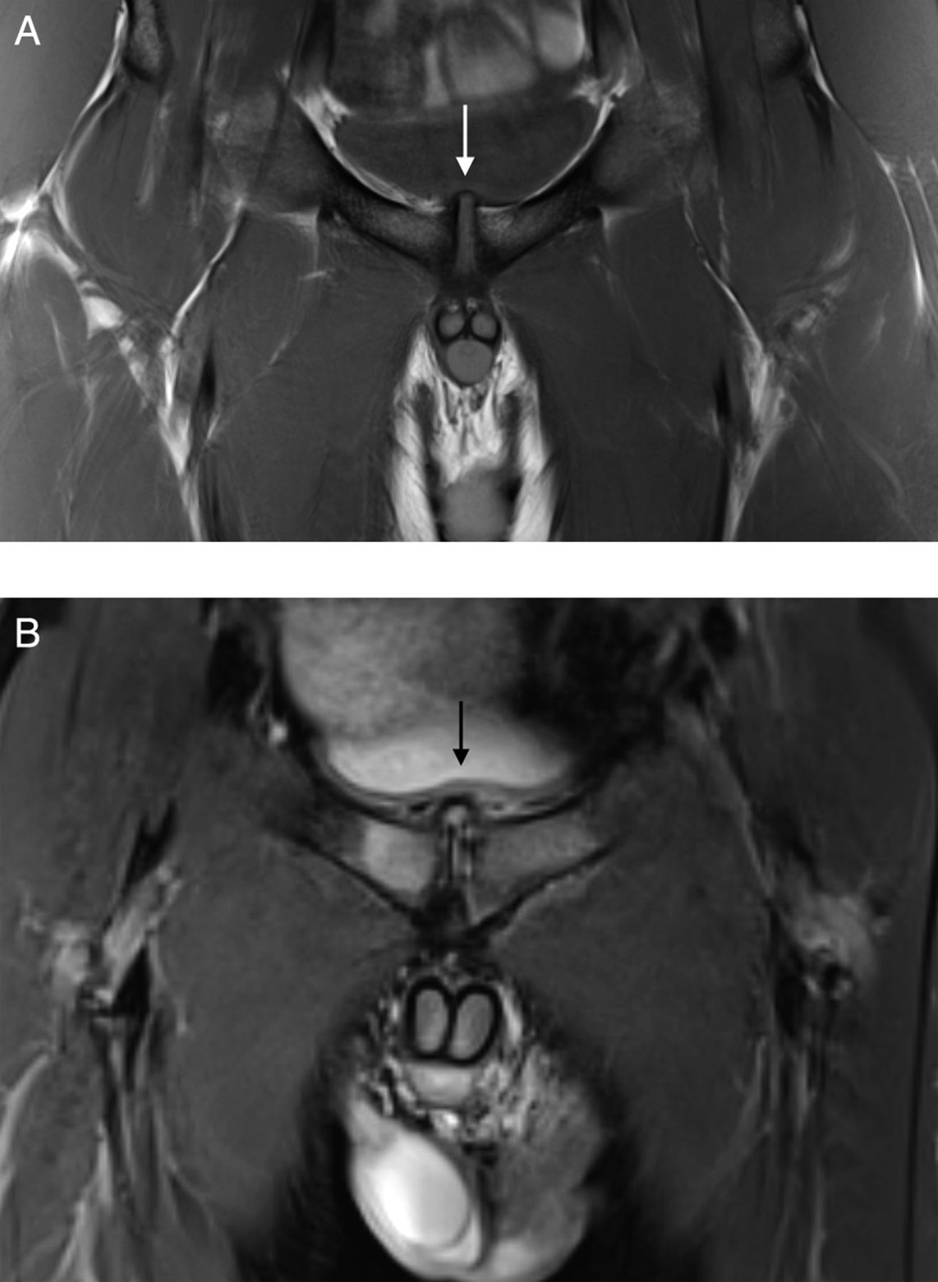
MRI evaluation protocol

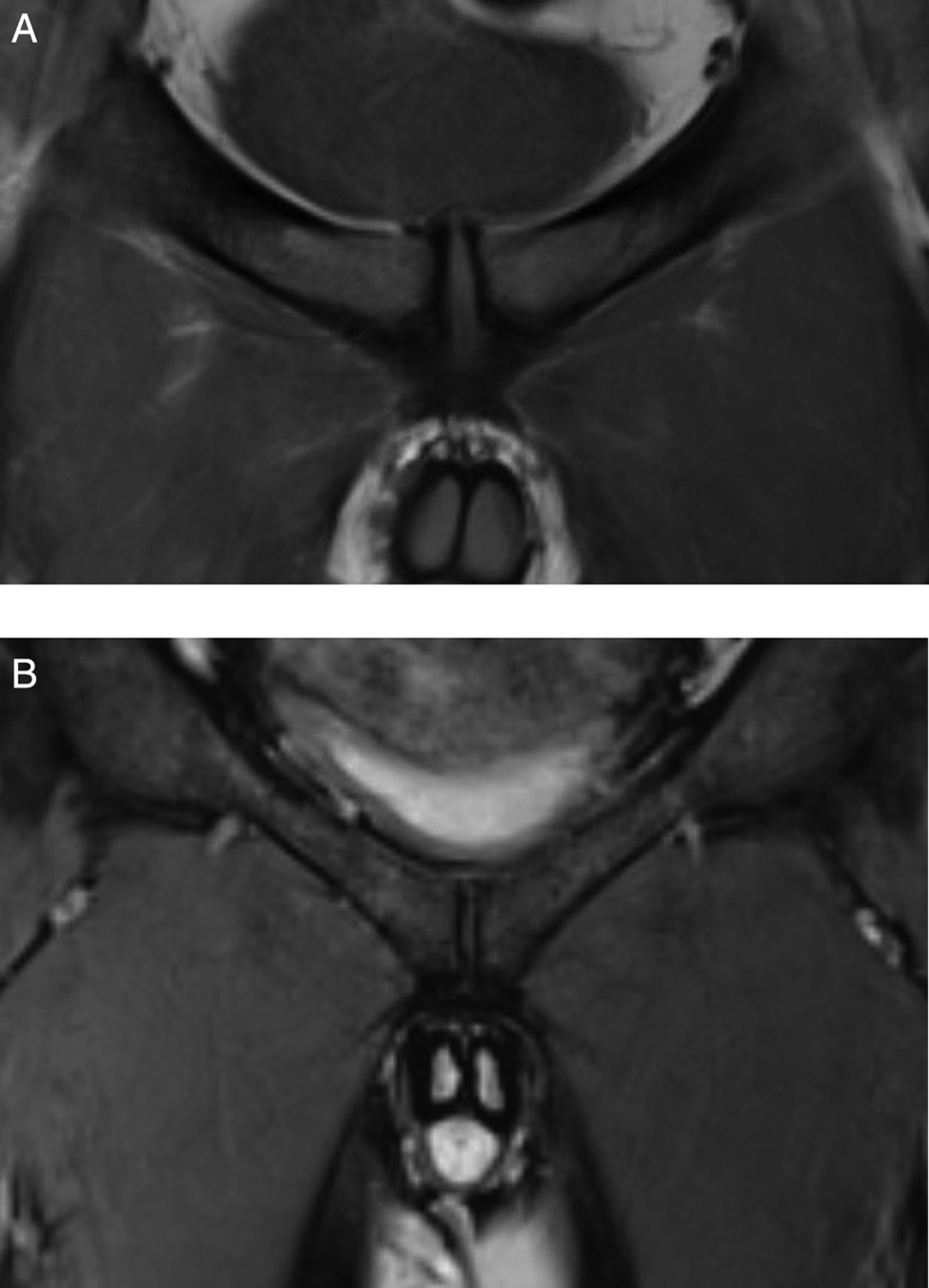
Coronal MRI view of the symphysis pubis—normal symphyseal joint (A)T1-weighted, (B) short tau inversion recovery (STIR).
Diffuse increased signal intensity within the pubic bone marrow (BMO) (A) on coronal STIR sequence, (B) on axial oblique T2-fat saturation (FatSat) sequence.

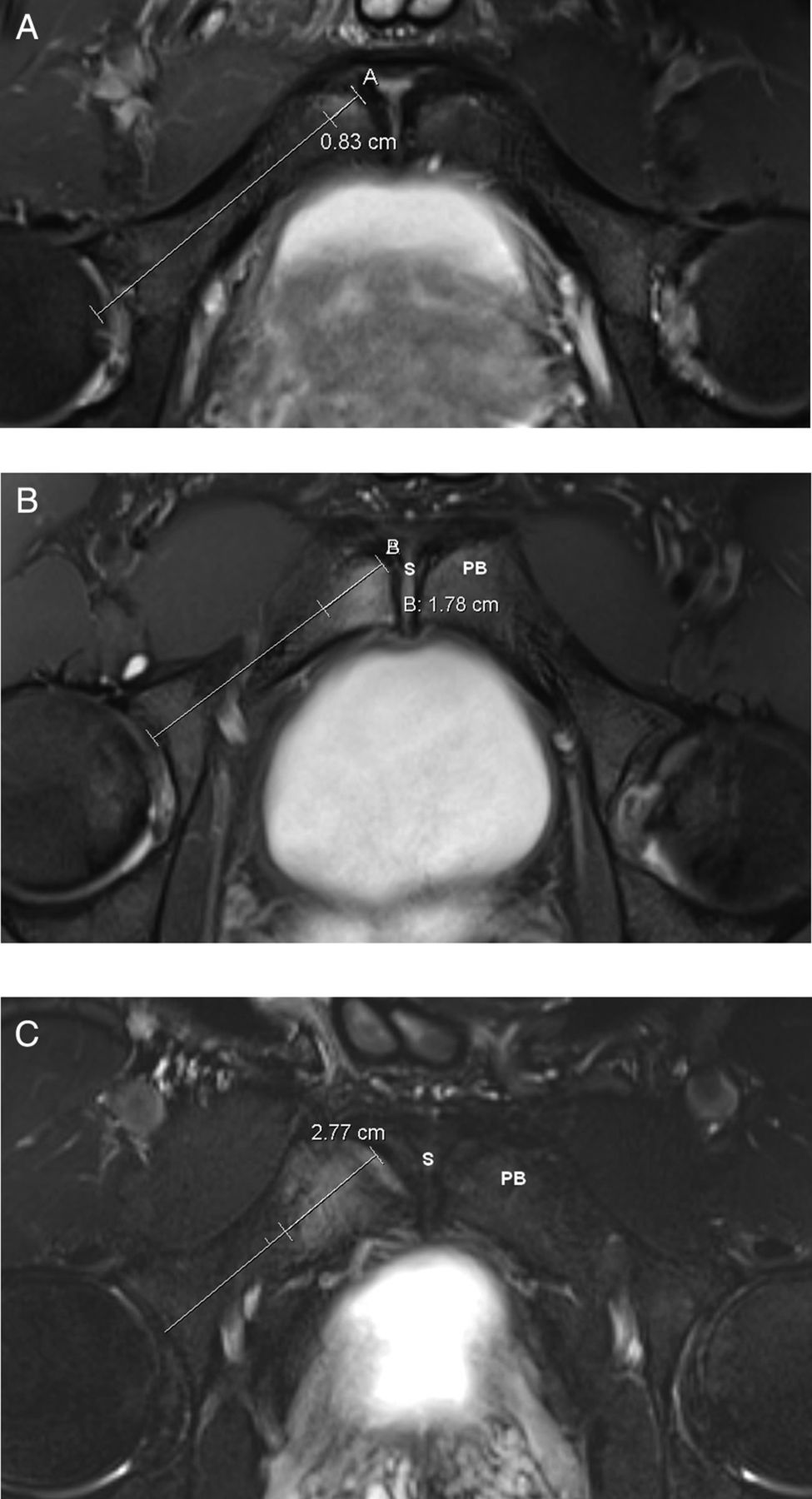
Pubic bone marrow oedema on fluid-sensitive axial oblique sequences (A) grade 1 extending less than 1 cm from symphysis along long axis of superior pubic ramus, (B) grade 2 extending more than 1 cm and less than 2 cm from symphysis, (C) grade 3 extending more than 2 cm from symphysis.
Statistical analysis
Pearson χ² statistics (or Fisher's exact test) and ORs with 95% CIs were calculated for each binary variable to detect any association between clinical ARGP and positive MRI findings on the corresponding side (right and left leg), when comparing symptomatic and asymptomatic soccer players. The ordinal variable BMO (grade 0–3) was evaluated by a χ² test for trend. Since MRI findings related to anatomical midline structures (eg, central disc protrusion) would be counted twice for the symptomatic and asymptomatic legs of players with ARGP, the 17 asymptomatic legs of players with ARGP were excluded from the analysis. The variable central disc protrusion was analysed on the individual participant level, and not according to the side/leg. Statistical tests were repeated in the same manner to compare asymptomatic and asymptomatic non-soccer players to detect any association between soccer play and positive MRI findings. A p value of less than 0.05 was considered statistically significant. No power analysis was applied as this study has an exploratory character. All analyses were performed with SPSS Statistics V.19.0.
Results
Characteristics of all study participants are displayed in table 4. Symptomatic soccer players reported symptoms lasting for a median of 42.5 weeks (range 4–624 weeks). Of the 28 soccer players with ARGP, 8 had left-sided pain, 9 had right-sided pain, and 11 had bilateral pain, representing 39 symptomatic adductor tendons/sides, and 17 asymptomatic sides that were excluded from statistical analysis. Both right and left legs/sides of control participants were evaluated: the 17 asymptomatic soccer players thus had 34 asymptomatics legs, and the 20 non-soccer playing controls had 40. Importantly, in this study, we recorded a significant difference in all HAGOS scores between symptomatic soccer players and asymptomatic athletes (p<0.001), indicating that our three groups of participants were well selected.
Characteristics of study participants (p values indicate comparison with asymptomatic soccer players)
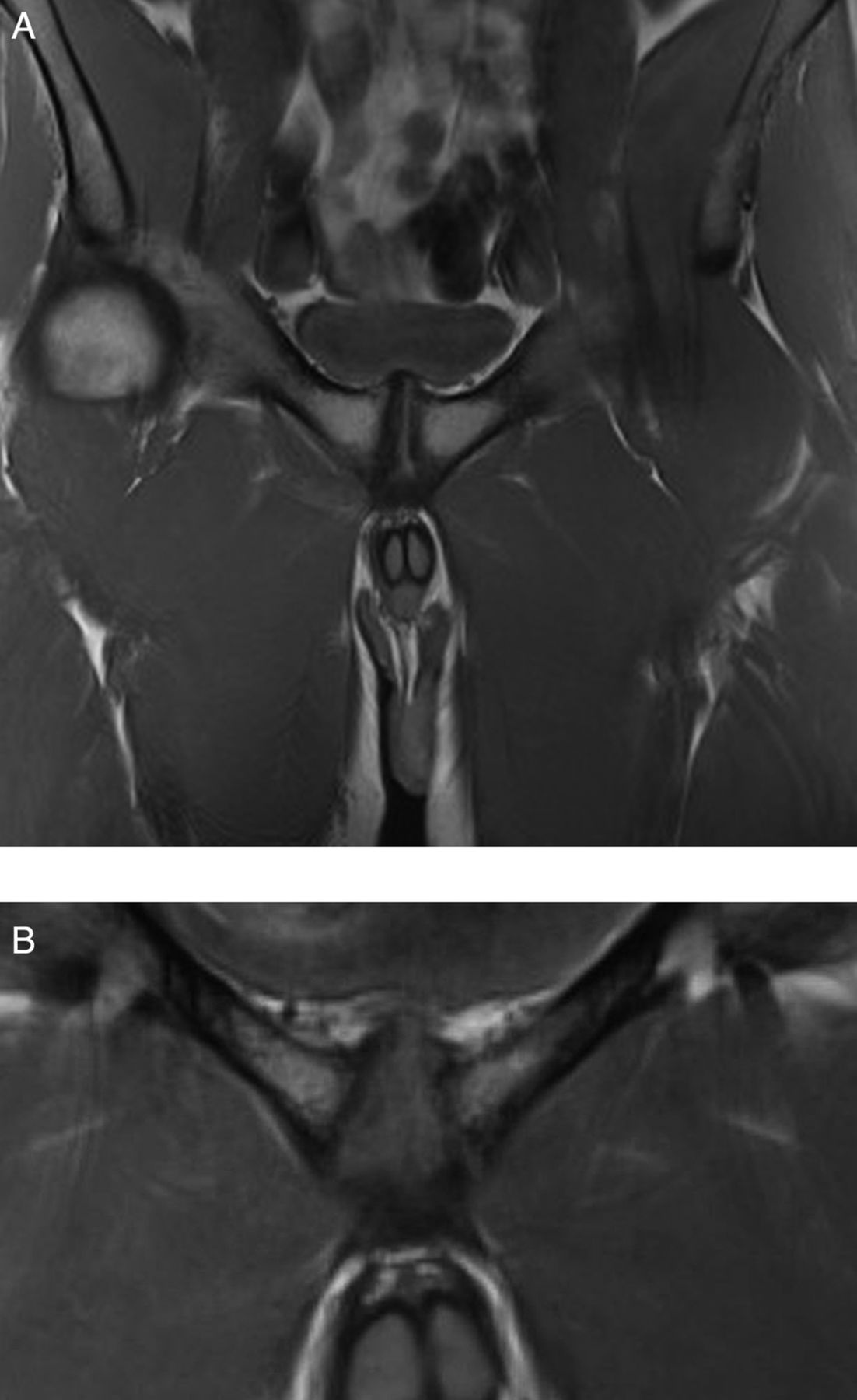
For the variable central disc protrusion, the 28 symptomatic players, 17 asymptomatic players and 20 non-soccer players were compared on the individual level. MRI findings and statistical analyses are displayed in tables 5 and 6. Although there was no association between the overall presence of BMO (grade>0) and ARGP in soccer players (p=0.11), there was a statistical trend (p=0.027) towards more severe grades of BMO in symptomatic players than in asymptomatic players. There was no significant association between ARGP in soccer players and any predefined MRI findings in the protocol, except for disc protrusion (p=0.027; figure 6A, B). Significantly higher odds (OR from 6.3 to 13.3) for the following findings were found in asymptomatic soccer players compared with asymptomatic non-soccer players: BMO, symphyseal sclerosis, parasymphyseal high-intensity line, subchondral cysts and joint surface irregularities, disc protrusion, and adductor tendinopathy (figure 7A–C). There was also a statistical trend towards more severe grades of BMO in asymptomatic soccer players compared with asymptomatic non-soccer players (χ² test for trend, p=0.045; table 6).
MRI findings counted per sides (left/right) in soccer players with adductor-related groin pain and 2 control groups
MRI findings

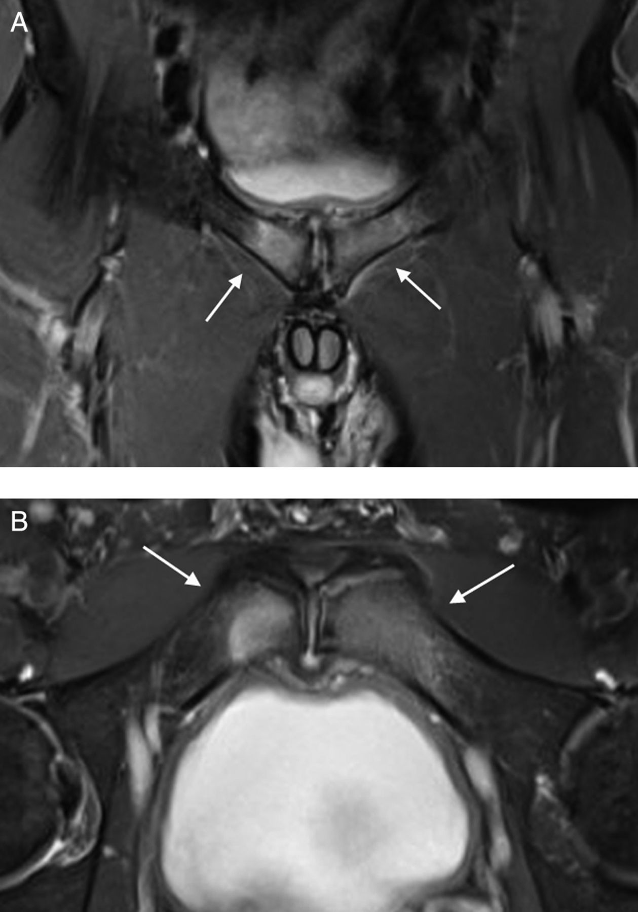
Central symphyseal disc protrusion (A) coronal T1-weighted, (B) coronal short tau inversion recovery (STIR).

Adductor longus tendon on Axial oblique fat saturation (FatSat) sequence (A) normal dark, well-delineated tendon in an asymptomatic non-soccer player, (B) adductor longus tendons display bulging and intratendinous increased signal intensity in an asymptomatic soccer player, (C) adductor longus tendinopathy with bulging and increased signal intensity within tendon in a soccer player with adductor-related groin pain.
Discussion
We wished to examine soccer players with and without ARGP and asymptomatic non-soccer playing athletes of matching age and activity to investigate whether pathological MRI findings, defined according to a standardised evaluation protocol, were associated with ARGP and/or soccer play. Overall, in our study, ARGP in soccer players was associated with central disc protrusion and more severe grades of BMO, but not with any other predefined pathological MRI findings. Interestingly, we detected statistically higher prevalences of positive MRI findings in asymptomatic soccer players compared with non-soccer players for the majority of MRI features.
MRI findings
BMO
In this study, soccer players with ARGP displayed significantly more severe grades of BMO than asymptomatic players (p=0.027), even if the overall presence of BMO of any grade in symptomatic players was not significantly higher than in asymptomatic players (p=0.11). The presence of BMO was significantly higher (p=0.007) in asymptomatic soccer players compared with non-soccer playing athletes. In line with our results, Verrall et al18 found an association between groin pain in Australian rules footballers and BMO extending more than 2 cm from the symphysis (p<0.01). Likewise, Lovell et al19 reported substantial BMO in 100% of symptomatic and 60% of asymptomatic soccer players, Paajanen et al16 observed more severe grades of BMO in athletes with groin pain than in controls (soccer and ice-hockey players), and recently Robinson et al34 recorded BMO in 62% of asymptomatic elite soccer players. Unlike the results of this study, Verrall et al18 reported no significant presence of BMO in asymptomatic athletes (footballers and runners). Cunningham et al35 reported BMO (described as either focal or diffuse, but not graded in severity) in 91% of soccer players with groin pain, but interestingly did not detect any pubic BMO in a mixed group of symptomatic and asymptomatic athletes (rowers and soccer players). The fact that our MRI study revealed BMO to be almost equally present in soccer players irrespective of symptom status, and much less prevalent in non-soccer players, combined with similar observations in several other MRI studies,16 ,18 ,19 strongly suggests that BMO can be present in many asymptomatic soccer players. BMO may reflect a sports-specific stress reaction in the pubic bones that sometimes becomes symptomatic, and the presence or absence of BMO does not discriminate between soccer players with and without ARGP. The presence of BMO may therefore only be a marker for regular football play, as only severe grades of BMO seem to be related to ARGP.
Degenerative changes at the symphyseal joint
Interestingly, we found a significantly higher prevalence of central disc protrusion in soccer players with ARGP, the significance of which is uncertain. The symphyseal joint shares important similarities with the intervertebral joints in the spine, as they are syndesmoses with a central disc, and with the adjacent bone marrow containing haematopoietic tissue.36 In the lumbar spine, Modic et al37 has described vertebral end plate changes with BMO (Modic type 1) followed by fatty infiltration (type 2) and potential fibrosis and sclerosis (type 3 and 4) in the context of progressive stages of degenerative spine disease.38 MRI-detected pubic bone marrow changes seem analogous to the Modic types 1–4 changes seen in the spine, as suggested by Gibbon and Schilders.39 Modic changes in the spine are located around the disc and vertebral end plates or at the site of entheseal attachment, such as the anterior and posterior longitudinal ligaments. In the symphyseal area, these changes could be related to the subchondral bone plate and/or the insertion of the inguinal ligament, rectus abdominis and adductor longus tendons, as these structures are all interconnected through their attachments to the pubic symphysis (joint capsule/ligaments). Modic changes are often combined with disc degeneration in the spine,37 ,40 and especially type 1 changes41–43 seem to be associated with clinical symptoms of low back pain.44 Interestingly, we recorded significantly more disc protrusion and severe grades of pubic BMO (equivalent to Modic type-1 changes) in soccer players with ARGP. All other MRI findings related to degenerative symphyseal changes were not significantly different in soccer players, whereas non-soccer players had significantly fewer degenerative changes (figure 8A, B). We observed fatty infiltration in the pubic bone marrow of 49% and 41% of symptomatic and asymptomatic soccer players, respectively, versus 5% of non-soccer players (figure 9A, B). Fatty infiltration has not previously been reported in MRI studies on groin pain in athletes, but it is reasonable to assume that it may correspond to a Modic type-2-equivalent middle stage of degenerative symphyseal joint disease, which could develop as a consequence of previous subcortical pubic bone oedema, indicating a previous stress response to excessive loading.45 Other MRI studies have recorded degenerative symphyseal changes in 20–98% of symptomatic athletes,16 ,18 ,30 ,35 ,46–48 and in 0–50% of asymptomatic athletes.16 ,18 ,30 ,35 Verrall et al18 and Paajanen et al16 found a higher proportion of pubic symphysis irregularities and subchondral cysts in athletes with current or past groin pain than in asymptomatic athletes, and Kunduracioglu et al48 reported a correlation of unspecified type and size (p<0.01) between symphyseal sclerosis and joint irregularities and chronicity of groin pain in soccer players, suggesting that symphyseal degenerative changes may to some extent be related to the duration of groin pain. We were, however, not able to show that degenerative findings of symphyseal sclerosis and subchondral cysts/joint surface irregularities were associated with ARGP in soccer players, but only that they were overall more present in soccer players than in non-soccer athletes. Our results suggest that soccer play itself may lead to the development of chronic degenerative changes at the symphysis reflecting excessive wear from soccer play. The fact that disc protrusion and high grades of BMO are more common in soccer players with ARGP than asymptomatic players suggests that ARGP may be related to these particular kinds of Modic-equivalent MRI findings at the symphyseal joint itself. Whether the pain experienced by the patient on palpation of the adductor tendon insertion and isometric contraction of the adductor muscle only originates from the adductor enthesis itself, or includes pain from traction or compression of the underlying pubic bone and symphyseal joint, needs further clarification.

Degenerative changes around the symphysis: central disc protrusion, joint surface irregularities, subchondral sclerosis, (A) coronal T1, (B) large view coronal T1.

Fatty infiltration around the symphysis appears (A) bright on coronal T1, and (B) dark on coronal short tau inversion recovery (STIR), respectively.
Pathology at the adductor muscle insertions
We anticipated higher prevalences of MRI findings related to the adductor tendon (adductor tendinopathy and musculotendinous lesions) in soccer players with ARGP than in asymptomatic players, but we found no such difference between the groups. However, MRI-defined adductor longus ‘tendinopathy’ was visible in 72% of symptomatic and 71% of asymptomatic soccer players, as opposed to 27% of non-soccer players, suggesting chronic structural changes in the adductor longus tendons of soccer players as a consequence of previous injuries, microtears and/or soccer-related overuse, irrespective of the current symptoms. These tendinous MRI changes, which we defined as increased signal intensity and/or thickening of the tendon (commonly referred to as tendinopathy49 ,50), do not seem to be related to symptoms, but rather suggest that our image acquisition protocol was not sensitive enough to discriminate between pathological symptomatic tendons and asymptomatic tendons in soccer players. Similar tendinous MRI changes have been shown in other tendons. Connor et al23 detected frequent MRI-detected rotator cuff tendinosis in the shoulders of asymptomatic overhead athletes. Gardin et al51 reported no correlation between tendon volume and pain symptoms in chronic Achilles tendinopathy, and Haims et al52 noticed that asymptomatic Achilles tendons frequently demonstrated increased intratendinous T2-weighted signal changes.
Three MRI studies evaluating adductor tendons in athletes with ARGP used intravenous gadolinium at 1.5 T: Schilders et al53 ,54 reported adductor enthesis contrast enhancement in 48% and 71% of clinically symptomatic athletes, but included no control groups for comparison. Robinson et al30 found a significant correlation between adductor enthesis enhancement and the symptomatic side of athletes with ARGP (r=0.37, p=0.008), which seems to suggest that the adductor longus enthesis is clinically implicated.
The MRI protocol used in this study was not able to discriminate whether changes in the adductor enthesis were related to symptoms or not. Injections of corticosteroid and local anaesthetic directed into the adductor enthesis53 ,54 have shown a potential for short-term alleviation of ARGP. Although this does not prove that the adductors are entirely responsible for the symptom complex ARGP, accumulating evidence suggests that the adductors play an important role in the development and existence of groin pain related to the pubic symphysis region.25 ,55–58 This is further supported by Holmich et al56 who showed that an active training programme specifically aimed at improving the strength and coordination of the adductor muscles is effective in treating ARGP. The symptom complex ARGP may therefore include the adductor longus tendon, the symphysis joint and the pubic bone, and the role of MRI in differentiating the possible pathologies in the adductor enthesis and tendons needs further investigation in future studies. The adductor tendon origins could be assessed using MRI scan protocols with thinner slices and smaller slice gaps, and possibly also by using intravenous gadolinium.30
Secondary cleft sign, superior cleft sign and parasymphyseal high-intensity line
Secondary cleft and superior cleft signs have been assimilated with adductor longus capsular lesions in other studies.35 ,46 ,59 ,60 We found secondary cleft signs (figure 10) in only four symptomatic and one asymptomatic soccer players, and only one secondary and one superior cleft sign in non-soccer playing athletes. Our study has therefore insufficient statistical power to suggest that these signs may not be related to clinically verified ARGP. Interestingly, Silvis et al61 observed that secondary cleft signs were present in 18% of asymptomatic ice hockey players. Parasymphyseal high-intensity lines paralleling the symphyseal subchondral bone plate resemble the secondary cleft sign (Branci, et al, 2014, unpublished work), but do not communicate with the symphyseal joint space. In this study, these findings were identified in soccer players (21% of symptomatic players and 24% of asymptomatic players) but in none of the non-soccer players, thus indicating a possible primary relation to soccer play itself.

Secondary cleft sign on coronal short tau inversion recovery (STIR) sequence. Curvilinear area with the signal intensity of fluid extending laterally and inferiorly from the inferior margin of the symphyseal joint, communicating with the symphyseal joint space.
Rectus abdominis tendinopathy
MRI-defined rectus abdominis ‘tendinopathy’ was rare in our cohort. This is in accordance with published studies reporting low prevalences of rectus abdominis tendinous injuries in soccer players.3 ,32 A previous MRI study59 reported rectus abdominis pathology in 65% of athletes suspected clinically of a combined rectus and adductor injury. As the adductor longus and rectus abdominis tendon insertions are located in close proximity,62 a diagnosis of rectus abdominis lesions in this study may possibly correspond to a diagnosis of adductor lesions in studies from other research groups, which could explain the difference in the prevalence of rectus abdominis lesions.
Strengths and limitations
As we have previously shown that the intraobserver and interobserver agreements were overall moderate when rating MRI findings according to our standardised evaluation protocol (S Branci, et al, 2014, unpublished work), and that the assessment of adductor longus tendinopathy was especially difficult to agree on, the MRI scans were consensus rated by three blinded radiologists in an attempt to optimise image interpretation. Our current image acquisition protocol did not enable a visual discrimination in rating adductor longus tendinopathy in symptomatic and asymptomatic soccer players, although we optimised our MR images with a high-field system (3 T) and a surface body coil. The cross-sectional design of the study does not allow us to conclude on cause–effect relationships between pathological MRI findings and ARGP symptoms. Our study would have benefited from larger study groups, as the prevalence of some of the MRI findings was low, but recruitment of especially asymptomatic soccer players was difficult due to lack of motivation for participating when players were injury free.
We included symptomatic soccer players with a well-defined clinical entity and controls that were comparable age-wise, sex-wise and activity-wise, differing only by their type of sports (soccer vs non-soccer athletes). No previous study has matched symptomatic and asymptomatic athletes in a blinded comparison, and in a manner that enables MRI findings to be associated with a specific sports-type irrespective of symptom status. As no previous studies have addressed this issue, our approach was of exploratory character with no power calculation. However, despite this limitation, our results clearly emphasise the importance of matching symptomatic athletes with activity-specific control groups, as we found significant differences in 6 of 11 specific MRI findings, suggesting a clear tendency of differences in imaging findings between asymptomatic soccer players and asymptomatic non-soccer players.
For evaluation of MRI findings related to the pubic symphysis, the pubic bones, and the adductor and rectus muscle insertions we used a new standardised MRI protocol developed by our research group: the Copenhagen Standardised MRI protocol.11a As with any clinical instrument, we are open to modifications of this protocol as the technology changes, and in response to new data, but we believe it is an excellent starting point from which to build a more uniform and scientific evaluation of MRI features in athletes with long-standing groin pain. It would enable different research groups worldwide to compare their radiological results, which has so far not been possible.11
Finally, MRI scans contain inherent image artefacts63 that may simulate pathology. The most prominent artefacts in the groin region are motion artefacts from the urinary bladder in the phase-encoding axis of the image,64 truncation artefacts in relation to tissue interfaces where there is an abrupt change in signal intensity,65 and magic angle artefacts66 visualised as increased intratendinous signal intensity on scan sequences with a short echo time in tendons oriented at an angle of approximately 55° to the main magnetic field.67 Even when taking these relevant MRI artefacts into account,64 ,67 MRI changes consistent with adductor longus tendinopathy and degenerative symphyseal joint changes seem to be a common feature in soccer players irrespective of symptoms, and may therefore reflect chronic structural changes in the tendinous and bony tissues induced by soccer play. However, assessment of adductor longus tendinopathy and rectus abdominis tendinopathy may in the future be improved by fine-tuning MRI scan acquisition protocols, such as using thinner slices and smaller slice gaps, possibly also by using intravenous gadolinium,30 and volumetric scan techniques.68
From a clinical perspective, the majority of positive MRI findings around the pubic symphysis in soccer players with ARGP lasting for more than 4 weeks seem to be related to soccer rather than pain symptoms, and many of these findings are therefore difficult to use for diagnostic workup. Findings relevant for long-standing adductor-related pain could be severe BMO and disc protrusion.
Conclusion
ARGP in soccer players was only associated with central disc protrusion and more severe grades of pubic BMO, and was interestingly not associated with the majority of predefined MRI findings around the pubic symphysis. Moreover, positive MRI findings were significantly more frequent in soccer players compared with non-soccer players irrespective of symptoms. Positive MRI findings may thus be associated with soccer play itself rather than ARGP, which has important implications for the diagnostic workup of these patients. Further development of MRI scan protocols is needed to clarify the significance of the adductors for the clinical entity ARGP.
What are the new findings
Adductor-related groin pain in soccer players was associated with central symphyseal disc protrusion and severe grades of pubic bone marrow oedema. Interestingly, the majority of commonly investigated MRI findings around the pubic symphysis in soccer players were not associated with adductor-related groin pain.
A high proportion of asymptomatic soccer players displayed positive MRI findings.
The majority of positive MRI findings were much more frequent in asymptomatic soccer players compared with asymptomatic non-soccer players.
Positive MRI findings may thus be associated with soccer play itself rather than existing adductor-related groin pain.
How might it impact on clinical practice in the near future
The fact that positive MRI findings may be associated with soccer play rather than clinical adductor-related groin pain has important implications for the diagnostic workup of these patients.
A standardised MRI evaluation protocol enables researchers to compare and reproduce MRI findings in published studies on pathological findings in athletes with hip and/or groin pain.
Further development of MRI scan acquisition protocols is needed to clarify the significance of the adductors for the clinical entity adductor-related groin pain.
Acknowledgments
The authors wish to thank Professor Carsten Thomsen and radiographer Poul Henrik Frandsen for their help with programming MRI sequences, Ulla BraschMogensen, Department of Biostatistics, Copenhagen University for statistical assistance, medical student Martin Nielsen for practical assistance, and the medical and physiotherapy students who helped with the initial contact with soccer coaches and teams.
References
Footnotes
Contributors SB contributed to planning the study, recruiting participants, MRI scanning participants, drafting and developing the MRI evaluation protocol, analysing images, analysing, interpreting and reporting statistical data, designing, writing and revising the manuscript. KT and PH contributed to planning the study, recruiting participants, examining participants, interpreting and reporting statistical data, designing, writing and revising the manuscript. BHB, MB and MBN contributed to planning the study, drafting and developing the MRI protocol, interpreting and analysing images, designing, writing and revising the manuscript.
Competing interests None.
Ethics approval The study was approved by the Danish National Committee on Health Research Ethics (H-2-2010-127), and the Danish Data Protection Agency (2011-41-5964).
Provenance and peer review Not commissioned; externally peer reviewed.