Article Text

Abstract
Objective The purpose of this study was to determine the prevalence of muscle imbalance among young adolescent acrobats (n=15) and if there was a potential link to injury.
Methods Isokinetic strength of the lower extremity, isometric strength of the trunk, and flexibility of both the trunk and lower extremity were assessed. Pearson correlation (r) and χ2 correlation tests were performed on all explanatory variables.
Results Significant correlations were found between isokinetic peak torque of the quadriceps and hamstrings (p=0.000) and the plantar flexors and dorsiflexors (p=0.000) on both sides, along with plantar flexor dominance (p=0.000). Non-significant findings were seen when identifying dominance between the quadriceps and hamstrings (p=0.933) as well as when correlating peak torque and flexibility of the lower extremity (right hamstrings: p=0.668, left hamstrings: p=0.338, right quadriceps: p=0.171, left quadriceps: p=0.707, right plantar flexors: p=0.282, left plantar flexors: p=0.382, right dorsiflexors: p=0.297 and left dorsiflexors: p=0.393).
Conclusion Acrobats demonstrated noticeably high ranges of flexibility, and the most common injury site was found to be the ankle. However, these mentioned injuries were not all due to acrobatic participation. The limited sample size warrants extensive research with a larger sample size to further verify or dispute the results found in this study. Muscle imbalances found within this population could increase the risk of injury.
- muscle imbalance
- injury
- isokinetics
- acrobats
- dance
Data availability statement
Data are available upon reasonable request.
This is an open access article distributed in accordance with the Creative Commons Attribution Non Commercial (CC BY-NC 4.0) license, which permits others to distribute, remix, adapt, build upon this work non-commercially, and license their derivative works on different terms, provided the original work is properly cited, appropriate credit is given, any changes made indicated, and the use is non-commercial. See: http://creativecommons.org/licenses/by-nc/4.0/.
Statistics from Altmetric.com
WHAT IS ALREADY KNOWN ON THIS TOPIC
Dancers require enhanced physiological adaptations. A significant amount of muscle tension is developed, increasing the risk of overuse injuries.
WHAT THIS STUDY ADDS
An increased range of motion could protect acrobats from injury. However, more research on the effect of injury risk caused by hypermobility and imbalances of flexibility would be of great benefit, particularly in acrobatic dance.
Quadriceps dominance along with plantar flexor dominance is noticeable among an acrobatic population.
HOW THIS STUDY MIGHT AFFECT RESEARCH, PRACTICE AND/OR POLICY
Research with the inclusion of exercise interventions focusing on holistic training programmes to reduce muscle imbalances and ultimately identify if the reduction in muscle imbalance influences injury prevalence is encouraged. Further data such as the recording of menarche in this population will also be insightful to see if there are any significant relationships between muscle imbalance, injury risk and menarche status.
Introduction
Acrobatics is a physically demanding sport that pushes an individual’s body to unusual limits to perform a variety of tricks that can be seen as unnatural movements. Through that, the body is used in unique ways. Acrobatic dance in South Africa is seen as a style that incorporates acrobatics and floor gymnastics with classical dance techniques and contortion.1 The execution of acrobatic skills holds a place in the history of the international gymnastic discipline,2 and this style can be seen on various stages and theatrical arenas.2
Previous studies have been conducted on athletes with similar performance demands, such as gymnasts, and an immense amount of data can be found among ballet dancers.3 Training for dancing, such as acrobatics, is unlike other sports, which have seasons, and is rather an annual endeavour. Due to this frequent duration, dancers’ risk of overuse injuries increases.4 Dancers require enhanced physiological adaptations due to the great physical demands of the sport, where both aerobic and anaerobic systems are heavily relied on, and a significant amount of muscle tension is developed.5 Multiple studies have found that the repetition of movements in a variety of sports leads to adaptations of different tissues including the muscles.6 According to Coombes and Garbutt,7 muscle imbalances and bilateral muscle asymmetry are the aetiology of many injuries due to the repetitive nature of dance8 and the annual training calendar. It would, therefore, be important to identify if acrobats, too, are at risk to muscle imbalance and possible injury aetiology.
A method in which muscle imbalance can be tested and identified is through isokinetic testing. The Hamstring:quadriceps peak torque ratio is a common influence on muscle imbalances associated with sport-related injuries.9 In a study done by Kuni and Schmitt,10 isokinetic testing found a balance deficit between the plantar flexors and dorsiflexors in professional dancers. Testing the isokinetic strength of the ankle joint of female acrobats would be useful to note if similarities to this study are present and if they possibly may be linked to injury onset.
In addition, it has been suggested that flexibility imbalances have a causal relationship with dance injuries.11 In a study done by Steinberg et al,12 42.6% of dancers they screened were injured, and one of the factors associated with the injuries seen was the range of motion, specifically the presence of either hyper or hypo range of motion.
Study objectives
This study aims to identify if there are muscle imbalances among female acrobats in South Africa and hopes to discover if there is a correlation to injury. This will be done through objective testing of their lower limb strength, their lower limb flexibility and the filling out of an injury history survey. This research study could provide insight into the factors that may cause injury in acrobats and shed light on the physiological condition of acrobats in South Africa, which has not been thoroughly investigated. Furthermore, this research study and its results could potentially provide interest in further investigation into this dance style and other dance styles.
Methods
Subjects
Female acrobats (n=15) volunteered to participate in the study. All participants were active training acrobats (hours trained weekly=2.67±0.7) that were uninjured, pupils of studios within Johannesburg in South Africa and satisfied with a physical activity readiness questionnaire. Acrobatic training normally consists of a warm-up and stretching for 15 min before practising routines and acrobatic tricks, which test the individuals’ strength and flexibility.
Procedures
Subjects visited the biokinetic clinic located on the University of Johannesburg’s campus to participate in one session where all data were collected. Height and weight measurements were obtained first, along with their age. Height and body mass were measured using a scale and a Seca stadiometer. Intermission or rest between each exercise test mentioned further was not set, but rather a continuous assessment was followed, only pausing at the participant’s request if needed.
Isokinetic testing protocol
Participants were warmed up before the isokinetic testing by cycling on a cycle ergometer for 5 min. Five familiarisation contractions were also carried before or to each movement tested. Test measurements were taken shortly after the familiarisation contractions when the participants stated they were ready. The isokinetic testing was performed using a Cybex Norm (division of Lumex, Ronkonkoma, Long Island, New York, USA). Calibration was performed each day before testing. The following two movement patterns were performed: seated knee flexion and extension and prone plantar and dorsiflexion (hip and knee in an extended position). The movement velocity was set at 60°/s for the knee and 30°/s for the ankle. Participants were positioned according to the standardised procedure described by Perrin.13 Five familiarisation repetitions (two repetitions at 50%, two repetitions at 75% and one at 100%) at increasing effort levels were carried out before the five maximal concentric contractions were performed for both movement patterns. Correction for gravity was made for both movement patterns. Participants were encouraged verbally to perform their best during both tests. Participants were also given visual feedback during the testing. Peak torque in newton-metres (Nm) was recorded as the highest of five repetitions, and the agonist:antagonist ratio (%) was calculated for both movement patterns.14
Back strength
Back strength was measured using the procedure outlined by Heyward and Gibson.15 A calibrated back–leg–chest (BLC) dynamometer was used to test the lower back strength of each participant. The chain length was adjusted according to the participants’ height. This was achieved by asking the subject to stand on the base of the BLC dynamometer with their knees extended, and the handle was positioned at the height of the distal thigh, just superior to the knee joint. For the test, the participants had to stand on the base with their knees extended, hips slightly flexed, chests out slightly, shoulders rolled back, and neck and back straight. The bar was held right-hand palms down and left hand holding the bar palms up. The participants were asked to try lifting the bar in a vertical direction through their lower back. After demonstration and familiarisation trials, three test trials were completed for an average to be calculated.
Forearm plank
Participants were asked to lie on a mat and assume a forearm plank position according to the procedure followed by Strand et al.16 They were instructed to maintain this position for as long as possible and verbal cues were given to encourage adherence to test validity. When the participant assumed the correct position, the stopwatch was started. The test was terminated when (1) the participant fatigued or voluntarily stopped; (2) the participant failed to maintain the correct position; and (3) the participant verbally expressed the ill effects of participation in the test. Participants were provided with verbal cues if their technique wavered from the desired position during the test. However, the test was terminated when two consecutive corrective cues were given with an inadequate correction in form. Each subject only performed the test once to keep consistent with other types of fatiguing fitness assessments.16
Lumbar flexion
Before participants performed the test, a rotational trunk stretch was conducted, and the movements of the trunk that were to be tested were excluded to prevent the practice effect.17 Lumbar flexion was measured using a measuring tape using the standardised method of the Modified–Modified Schober test.17 The participants were instructed to stand erect, with their arms at their sides, and their feet were placed on two marked spots 15 cm apart. The movement was shown to the participant by the tester, ensuring they knew to keep their arms hanging in front and to keep their knees straight. Once the participant understood what was meant to be conducted, the tester kneeled behind the standing participant and identified both posterior superior iliac crests and marked them. The midpoint between those two marks was measured and marked (inferior marking). Lastly, 15 cm was measured up the spine and marked (superior marking). The tester aligned the measuring tape between the two skin marks, 0 cm being on the inferior marking and 15 cm being on the superior marking. The measuring tape was kept securely on the participants’ back while they bent forward as far as possible while keeping their knees straight. The distance from the inferior marking to the superior marking was then recorded. The range of motion was the difference between 15 cm and the length measured at the end of motion. This procedure was performed three times for an average to be calculated, and after each measurement, the participant was told to ‘relax and come into a comfortable standing position’.
Lumbar extension
The same landmarks used for the lumbar flexion test were used to measure lumbar extension. The participants were instructed to stand erect, with their arms at their sides, hands placed on their buttocks and their feet placed on two marked spots 15 cm apart. The movement was shown to the participant by the tester, ensuring they knew to keep their knees straight and their hands on their buttocks while they performed lumbar extension. The tester aligned the measuring tape between the two skin marks, 0 cm being on the inferior marking and 15 cm being on the superior marking. The measuring tape was kept securely on the participants back while the participant bent backwards into full lumbar extension, and the new distance between the two markings was measured. The difference in distance between the two marks was used to indicate the amount of range of motion. This procedure was performed three times for an average to be calculated, and after each measurement, the participant was told to relax and come into a comfortable standing position.
Lumbar lateral flexion
Before the test was conducted, participants were asked to warm up with three repetitions of rotational back stretches, excluding stretching in planes that were to be tested to prevent a practice effect.17 The procedure that was followed mimicked the test performed by Malik et al,17 and a flexible fibreglass tape measure of 90 cm was used.
Standard sit and reach
The standardised procedures found in the ACSM’s Guidelines for Exercise Testing and Prescription18 were used for testing. Each participant was asked to remove their shoes and sit with their feet placed flat on the sit-and-reach box on the two marked spots 15.2 cm apart. The participants were then instructed to place their hands on top of each other and reach as far forward as possible without bending their legs. They were also told to exhale and drop their heads between their arms when reaching forward. This test was conducted three times, and an average was calculated.
Goniometry
The hamstrings and quadriceps range of motion was measured according to the guidelines set out by Clarkson19 and Palmer and Epler.20 A baseline goniometer of 15 cm was used, and each movement was repeated three times on each side to calculate an average.
The plantar flexors and dorsiflexors range of motion was measured by having the participants lie supine with their ankles placed off the plinth to ensure the plinth did not restrict movement. The goniometer fulcrum was placed on the lateral malleolus. The goniometer’s immovable and movable arms were placed in line with the fifth metatarsal with the ankle in a neutral position. As the participant performed each movement (dorsiflexion and plantarflexion), the immovable arm stayed fixed in the starting position (neutral), and the movable arm was moved to be in line with the fifth metatarsal at the end of the participants range of motion in both movements (dorsiflexion and plantarflexion). A baseline goniometer of 15 cm was used, and each movement was repeated three times on each side to calculate an average.
Injury survey
No specific survey for acrobatic dance injuries was found. As such, the SafeDance IV survey report21 was used as a foundation and modified for the context of this study. An online survey was sent to each participant to fill in to gather information on injury history. The survey detailed the dancer’s demographics, the number of hours trained weekly, any musculoskeletal injuries experienced including the site of injury, type of injury and if the injury was caused by acrobatic training. The definition of injury for this investigation was ‘any body part that has been hurt and interferes with your acrobatic training’.12 It was also specified that if they experienced more than one injury, they were to report the injury that most affected their acrobatic training and dance performance. Each individual’s injury survey was completed straight after the physical testing session.
Data analysis
Descriptive and inferential statistical analyses were used to analyse the data collected. Pearson correlation (r) tests were performed on all explanatory variables to test for any relationships present with muscle imbalance or injury. χ2 tests were also conducted to substantiate the results further. SPSS V.27.0 was used for all statistical analyses. The statistical significance was set at 95% (p<0.05).
Results
The following results of the female acrobats’ (age=13.60±2.29, height=1.58±0.09 m, body mass=51.37±12.92 kg) lower limb strength, lower limb flexibility and injury history were found.
Figure 1A displays a correlation graph that compares the Isokinetic peak torque (Nm) of the right hamstrings and right quadriceps. There was a highly significant, positive linear correlation between the isokinetic peak torque of the right hamstrings and the isokinetic peak torque of the right quadriceps (r=0.848, p=0.000). There was also a significant, positive linear correlation for the left side’s hamstrings and quadriceps peak torque values (r=0.943, p=0.000), as shown in figure 1B.

(A) Scatterplot depicting the correlation between right isokinetic peak torque of the quadriceps and right isokinetic peak torque of the hamstrings. (B) Scatterplot depicting the correlation between left isokinetic peak torque of the quadriceps and left isokinetic peak torque of the hamstrings.
The prevalence of hamstring or quadriceps dominance in the acrobats was also tested. Of the 15 acrobats tested, 9 were quadriceps dominant on both sides. However, the limited sample size of this study group did not lead to any significant findings (p=0.933). Similarly, the prevalence of dorsiflexor or plantar flexor dominance was tested. Out of the 15 acrobats tested, 14 were plantar flexor dominant on both sides. This was confirmed by the low p value (0.000). However, due to the limited sample size of this study group, whether these relationships will exist in larger sample groups cannot be assumed.
Figure 2A displays a correlation graph that compares the isokinetic peak torque (Nm) of the right and left plantar flexors. There was a highly significant, positive correlation between the isokinetic peak torque of the plantar flexors (r=0.913, p=0.000). Figure 2B displays a correlation graph that compares the Isokinetic peak torque (Nm) of both the right and left dorsiflexors. There was a highly significant, positive correlation between the isokinetic peak torque of the dorsiflexors (r=0.951, p=0.000).
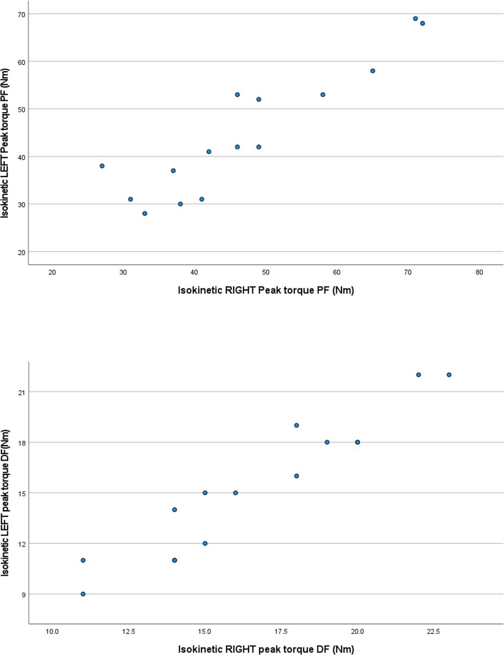
(A) Scatterplot depicting the correlation between left isokinetic peak torque of the PFs and right isokinetic peak torque of the PFs; (B). Scatterplot depicting the correlation between left isokinetic peak torque of the DFs and right isokinetic peak torque of the DFs. DF, dorsiflexor; PF, plantar flexor.
When correlating dorsiflexor isokinetic peak torque with plantar flexor isokinetic peak torque, significant findings were found for both sides as seen in figure 3A,B (right: r=0.670, p=0.006, and left: r=0.648, p=0.009). Physiological profile means were also calculated for a number of the tests performed (back strength=44.9±23.24 kg, forearm plank=50.65±12.31 s/ms, lumbar flexion=7.01±1.59 cm, lumbar extension=5.21±1.45 cm and sit and reach=52.79±8.52 cm).

(A) Scatterplot depicting the correlation between right isokinetic peak torque of the DFs and right isokinetic peak torque of the PFs (B). Scatterplot depicting the correlation between left isokinetic peak torque of the DFs and left isokinetic peak torque of the PFs. DF, dorsiflexor; PF, plantar flexor.
When analysing the results of trunk lateral flexion, the following observations were found. Participants with right-hand dominance, on average, had reduced flexibility in lateral trunk flexion when testing the right side’s flexibility (23.13±5.57 cm). In contrast, on average, participants with left-hand dominance performed better in the flexibility testing of lateral flexion of the right side (26±3.46 cm) (the opposite was true for the other side). The right-hand dominant individuals performed better on average in the lateral flexion testing of the left side (25.81±4.31 cm) in comparison to the left-hand dominant individuals (24.78±0.56 cm).
It can be seen in figure 4A,B that there was a non-significant positive correlation between hamstring peak torque and hamstring goniometer flexibility on the right side (r=0.121, p=0.668) and on the left side (r=0.266, p=0.338).

(A) Scatterplot depicting the correlation between the flexibility of right hamstrings and right isokinetic peak torque of the hamstrings; (B) Scatterplot depicting the correlation between the flexibility of left hamstrings and left isokinetic peak torque of the hamstrings.
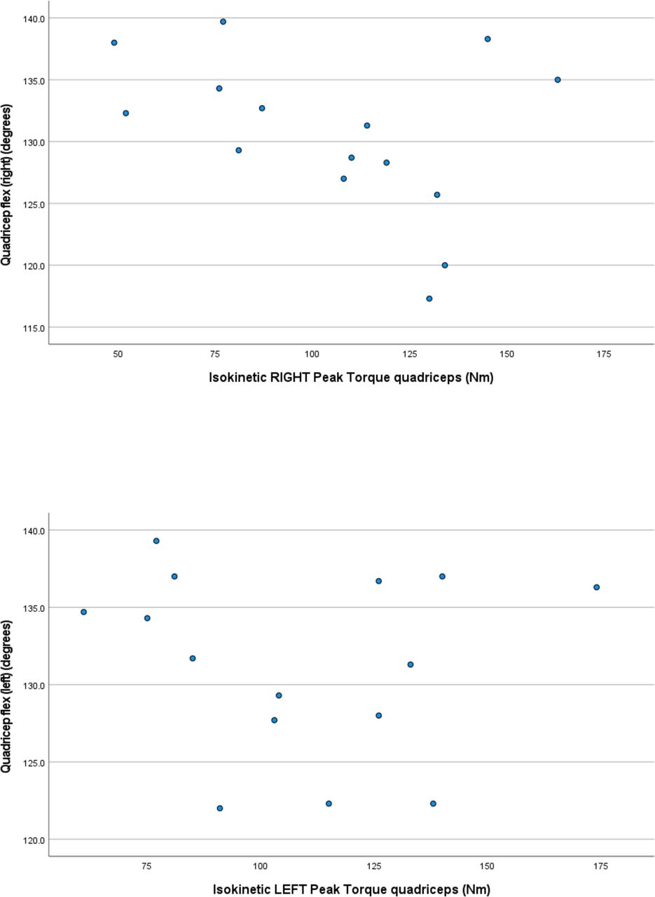
In figure 5A,B, it was also seen that there was a non-significant negative correlation between quadriceps peak torque and quadriceps goniometer flexibility on the right side (r=−0.373, p=0.171) and on the left side (r=−0.106, p=0.707). Similar findings were found with the testing of the plantar flexors and dorsiflexors. There was a non-significant positive correlation between the plantar flexor peak torque and plantar flexor goniometer flexibility on the right (r=0.297, p=0.282) and a non-significant negative correlation on the left (r=−0.244, p=0.382). There was a non-significant negative correlation between the dorsiflexor peak torque and dorsiflexor goniometer flexibility on the right side (r=−0.289, p=0.297) and left side (r=−0.238, p=0.393).
(A) Scatterplot depicting the correlation between the flexibility of the right quadriceps and right isokinetic peak torque of the quadriceps; (B). Scatterplot depicting the correlation between the flexibility of the left quadriceps and left isokinetic peak torque of the quadriceps.
Lastly, it was seen in the completed injury survey’s that the ankle was the most injured joint, with 5 of 15 participants (33.3%) reporting an ankle injury, with the knee being the second most injured joint (26.7%) of the participants. However, it is important to mention that these injuries did not exclusively occur during an acrobatic class or competition. Two out of the five ankle injuries occurred during acrobatic participation, and only one of the four knee injuries occurred during acrobatic participation, along with one back injury (26.7%). The remaining injuries mentioned in table 1 occurred in a different setting (73.3%).
Participant’s previous injury location and where the injury was experienced
Discussion
This study provides insight into the physiological profile of young female acrobats in Johannesburg, South Africa. Acrobats’ agonist and antagonist muscle groups tested via the Cybex Norm machine (quadriceps–hamstrings and plantar flexors–dorsiflexors) were seen to have a similar trend in all individuals. This observed trend strongly suggested that as an acrobat’s agonist peak torque increased, so would their antagonist peak torque. Despite the results being non-significant, it is still important to note that most of the acrobatic dancers were quadriceps and plantar flexor dominant. Quadriceps dominance was also seen in a study among trained dancers by Vogelpohl et al.22 It could be due to dancers’ reliance on classical dance movements that include knee extension and fewer movements incorporating knee flexion. Quadriceps dominance also suggests reduced hamstring strength compared with quadriceps strength and is implicated in increasing the risk for lower extremity injuries.23 Plantar flexor dominance may be a result of the dancers spending a lot of time on their toes in extreme plantarflexion,24 such as in relevé (action of standing on the ball of your foot), performing tricks and dance movements. Furthermore, plantar flexor dominance suggests a greater plantar flexor strength in comparison to the dorsiflexors. According to Fong et al,25 this increased plantar flexion strength can be a risk factor for an ankle injury, specifically sprains.
Dancers need to have sufficient lower limb muscle strength and adequate trunk stabilisation strength as well,26 which justifies the need for testing sufficient back and abdominal strength. No previous literature on reference values for forearm plank duration and lower back strength in dancer-specific populations could be found. However, a study conducted by Strand et al16 among college students found that the average forearm plank duration for female students was 83±63 s. Compared with the current study, the acrobats’ average forearm plank was noticeably below this value. It could be deduced that this was due to the age of the participants. Lastly, the average back strength results of the acrobats in the current study were 44.9±23.24. When compared with adolescents of a similar age group in a study conducted by Ten Hoor et al,27 it was found that the acrobats had a lower back strength result overall. The acrobats falling below for both these measures could warrant further research into the trunk stabilisation abilities of acrobats and if their lack thereof may affect their performance or increase their risk of injury.
Dancers require extreme ranges of motion to prevent injury and achieve their sport’s demands.11 According to Ruggieri and Costa,9 ballet dancers were reported to have a sit-and-reach flexibility of 22.8±4.1 cm. Gymnasts were reported to have sit-and-reach flexibility of 36.4±4.2 cm, indicating that the acrobats in this current study had a noticeably greater sit-and-reach flexibility than ballet dancers and gymnasts. However, it is important to note that the SD of the acrobats’ results was larger than the other two dance styles, showing more variation in the results. Lateral trunk flexion flexibility results were also seen to be higher on the acrobats’ non-dominant sides compared with their dominant sides. Opposite findings were true for flexibility and dominance in a study conducted by Neto et al28 on healthy subjects. Increased ranges of motion within a joint reduce muscle injury risk.11 However, hypermobility and flexibility imbalances between muscle groups could be associated with higher injury risk.29 It would also benefit from more research, particularly in acrobatic dance.
As previously mentioned, sit-and-reach flexibility was noticeably better than other dance styles, and it could be speculated that their lumbar forward flexion could be of a similar result as the movements are similar and recruit the same musculature.30 Acrobats perform various tricks that require extreme ranges of back extensions, such as backbends, forward walkovers and backward walkovers31; this could suggest why their trunk extension flexibility results were relatively high compared with a normal population.17 According to a study conducted by McKay et al,32 the acrobats’ quadriceps flexibility fell below the norm of their age group, suggesting possible tightness in their quadriceps. Following further flexibility, it was seen that there were no significant findings between the acrobats’ muscle peak torque and their muscle flexibility. It could be speculated that greater strength on one side most likely will not influence that same side’s flexibility.33 A study conducted by Agopyan further supported this finding,34 in which similar findings within hamstring isokinetic results and hamstring goniometry results were found.
Out of the limited sample size that this study tested, only seven participants experienced a dance-related injury. Only three of those injuries occurred in an acrobatic setting. The most common injury site among acrobats was the ankle, with the knee following closely. This is in contrast to what was found in the literature that reported that knee injuries were the most common injury, with the ankle being far less.35 These findings could suggest that even with possible muscle imbalances among the acrobats, it cannot be concluded that it was a result of acrobatic training alone.
A consensus could be reached that female dancers experience a significant number of injuries,9 specifically of the lower limb,2 and female acrobats in South Africa are seen to possibly show a similar finding. However, it must be noted that further investigation with larger sample size groups of injury prevalence in acrobatic dancers is needed before decisive claims can be made. These injuries have been seen to have a variety of possible causes, including muscle imbalance in strength7 and range of motion.11 Well-rounded exercise training programmes could aid acrobats in meeting the physiological demands of their sport by reducing muscle imbalance in strength and flexibility and consequently may aid in reducing injury prevalence.
Limitations
Due to the limited sample size within this study, the findings cannot be generalised to larger populations. This sample size indicates the limited number and availability of registered studios to approach in South Africa in conjunction with fluctuating COVID-19 restrictions. The lack of research conducted among acrobats and dancers in specific tests carried out in the study also added to the limitations. This also hindered the ability to refer and relate the current study’s results to research previously conducted. Furthermore, the lack of injury data in this study also contributes to the limitations of this study and its ability to correlate the prevalence of muscle imbalance to injury risk.
Conclusion
Quadriceps dominance along with plantar flexor dominance was seen within this limited acrobatic sample population. Dancers (particularly with gymnastics and ballet) are at risk of lower extremity injuries, and these muscle imbalances noticeable in the acrobatic community could potentially be seen as risk factors for lower extremity injury with further research required. Increased ranges of motion in sit-and-reach results were noted in this study of acrobats compared with other dance styles. This increased range of motion could protect the acrobats from injury. However, more research on the effect of injury risk caused by hypermobility and imbalances of flexibility would be of great benefit, particularly in acrobatic dance. No significant findings were found between the acrobats’ muscle peak torque and their muscle flexibility. It could be suggested that greater strength on one side most likely will not influence flexibility on the same side. Due to the limited sample size of the current study, few significant findings were reported regarding muscle imbalance and its potential influence on an injury. Despite this, new insight was added to the physiological make-up of young acrobats in South Africa, which has not been previously conducted and could potentially encourage more research to be spent on this population along with less researched dance styles. Research with the inclusion of exercise interventions focusing on holistic training programmes to reduce muscle imbalances and ultimately identify if the reduction in muscle imbalance influences injury prevalence could be extremely insightful and should be encouraged.
Data availability statement
Data are available upon reasonable request.
Ethics statements
Patient consent for publication
Ethics approval
This study involves human participants and was approved by the Faculty of Health Sciences Research Ethics Committee (REC-879-2021) at the University of Johannesburg. The participants gave informed consent to participate in the study before taking part.Informed consent, child assent as well as a Physical Activity Readiness Questionnaire+ form was signed and obtained before starting the physical testing.
References
Footnotes
Twitter @Habib_Noorbhai
Contributors DS was responsible for the conception and design of the study, data collection and drafting of the manuscript. HN assisted with data analysis, provided critical revisions, provided guidance to DS during the project and serves as guarantor for the study. All authors approved the final version and are accountable for all aspects of the work.
Funding The authors have not declared a specific grant for this research from any funding agency in the public, commercial or not-for-profit sectors.
Competing interests None declared.
Patient and public involvement Patients and/or the public were not involved in the design, conduct, reporting, dissemination plans of this research.
Provenance and peer review Not commissioned; externally peer reviewed.